Copyright
©The Author(s) 2019.
World J Clin Cases. Jun 6, 2019; 7(11): 1337-1343
Published online Jun 6, 2019. doi: 10.12998/wjcc.v7.i11.1337
Published online Jun 6, 2019. doi: 10.12998/wjcc.v7.i11.1337
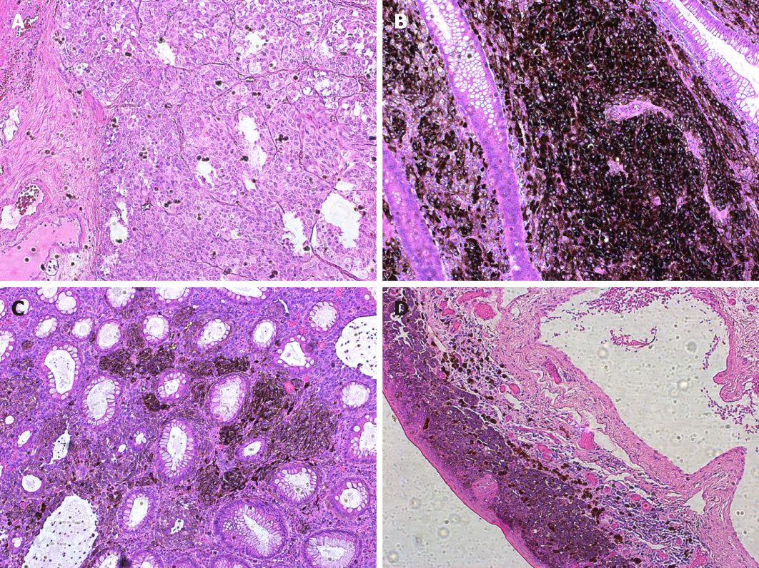
Figure 4 Histologic illustration of the four lesions in this case (HE staining, 10×).
A: Pedunculated mass located at the anal canal level. Although this lesion appeared unpigmented via visualization, scattered round atypical pigmented cells were found microscopically; B: The sessile mass 3 cm above the dentate line showed densely distributed pigmented cells; C and D: Two mucosal melanic zones were analyzed. Infiltration of atypical pigmented cells was found distributed in the mucosal and submucosal layers.
- Citation: Cai YT, Cao LC, Zhu CF, Zhao F, Tian BX, Guo SY. Multiple synchronous anorectal melanomas with different colors: A case report. World J Clin Cases 2019; 7(11): 1337-1343
- URL: https://www.wjgnet.com/2307-8960/full/v7/i11/1337.htm
- DOI: https://dx.doi.org/10.12998/wjcc.v7.i11.1337


