Copyright
©The Author(s) 2019.
World J Clin Cases. Jun 6, 2019; 7(11): 1337-1343
Published online Jun 6, 2019. doi: 10.12998/wjcc.v7.i11.1337
Published online Jun 6, 2019. doi: 10.12998/wjcc.v7.i11.1337
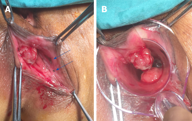
Figure 3 Transanal exploration of anorectal masses.
A: Derived from the anterior wall of the rectum, one pedunculated mass appeared at the anal canal level without melanin pigmentation. Two mucosal melanic zones were found at the anal canal level (blue arrows); B: Another pigmented mass was 3 cm above the dentate line under anoscope vision.
- Citation: Cai YT, Cao LC, Zhu CF, Zhao F, Tian BX, Guo SY. Multiple synchronous anorectal melanomas with different colors: A case report. World J Clin Cases 2019; 7(11): 1337-1343
- URL: https://www.wjgnet.com/2307-8960/full/v7/i11/1337.htm
- DOI: https://dx.doi.org/10.12998/wjcc.v7.i11.1337


