Copyright
©The Author(s) 2019.
World J Clin Cases. Jun 6, 2019; 7(11): 1315-1322
Published online Jun 6, 2019. doi: 10.12998/wjcc.v7.i11.1315
Published online Jun 6, 2019. doi: 10.12998/wjcc.v7.i11.1315
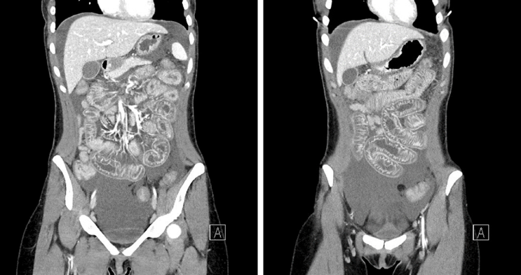
Figure 2 Computed tomography scan of the abdomen and pelvis with contrast, coronal view.
There is predominantly submucosal thickening/edema in the mid abdomen, thickened small bowel loops that are minimally dilated, no transition point, and moderate ascites.
- Citation: Gonzalez A, Wadhwa V, Salomon F, Kaur J, Castro FJ. Lupus enteritis as the only active manifestation of systemic lupus erythematosus: A case report. World J Clin Cases 2019; 7(11): 1315-1322
- URL: https://www.wjgnet.com/2307-8960/full/v7/i11/1315.htm
- DOI: https://dx.doi.org/10.12998/wjcc.v7.i11.1315


