Copyright
©The Author(s) 2019.
World J Clin Cases. Jun 6, 2019; 7(11): 1282-1290
Published online Jun 6, 2019. doi: 10.12998/wjcc.v7.i11.1282
Published online Jun 6, 2019. doi: 10.12998/wjcc.v7.i11.1282
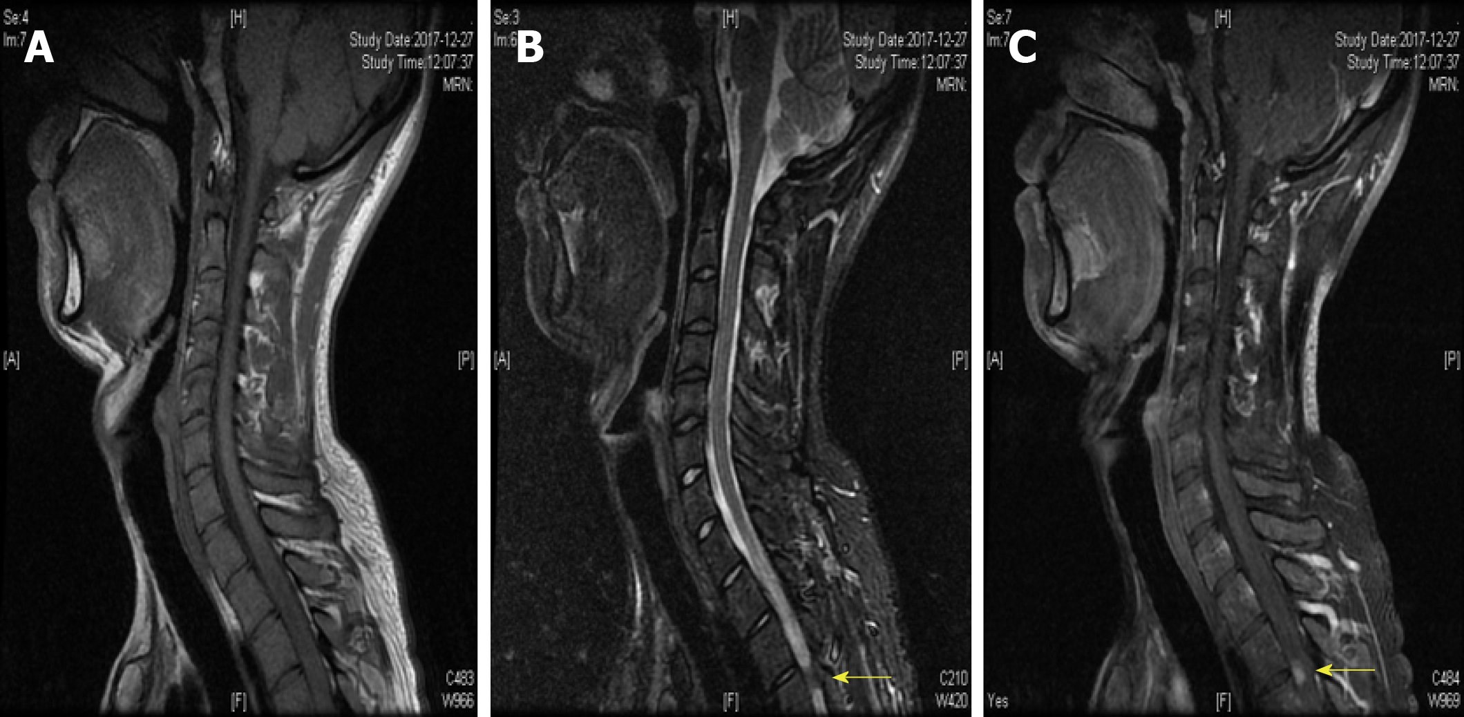
Figure 1 Spinal cord magnetic resonance imaging showed abnormal longitudinally extensive T2 weighted hyperintensities involving the posterior columns from C7 through T6, with "flip-flop sign" on cervical spinal magnetic resonance imaging.
- Citation: Yuan JL, Wang WX, Hu WL. Clinical features of syphilitic myelitis with longitudinally extensive myelopathy on spinal magnetic resonance imaging. World J Clin Cases 2019; 7(11): 1282-1290
- URL: https://www.wjgnet.com/2307-8960/full/v7/i11/1282.htm
- DOI: https://dx.doi.org/10.12998/wjcc.v7.i11.1282


