Copyright
©The Author(s) 2019.
World J Clin Cases. May 26, 2019; 7(10): 1234-1241
Published online May 26, 2019. doi: 10.12998/wjcc.v7.i10.1234
Published online May 26, 2019. doi: 10.12998/wjcc.v7.i10.1234
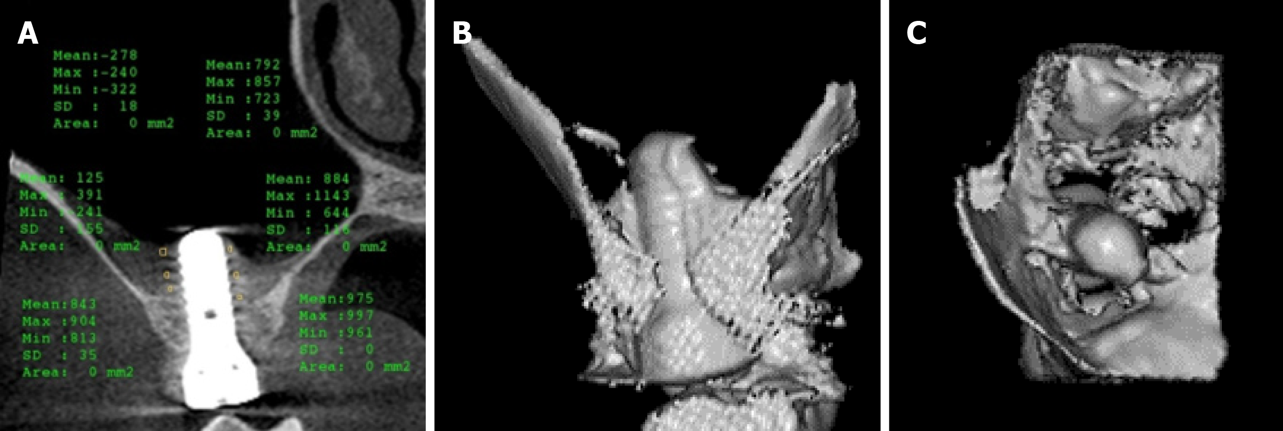
Figure 5 Cone-beam computed tomography images obtained at the 3-mo follow-up.
A: Coronal cone-beam computed tomography (CBCT) image showing the gained bone height around the implant; B: 3D CBCT image showing the gained bone from the buccal side; C: Another intra-maxillary 3D CBCT image showing the bone formation level at the same time.
- Citation: Mudalal M, Sun XL, Li X, Fang J, Qi ML, Wang J, Du LY, Zhou YM. Minimally invasive endoscopic maxillary sinus lifting and immediate implant placement: A case report. World J Clin Cases 2019; 7(10): 1234-1241
- URL: https://www.wjgnet.com/2307-8960/full/v7/i10/1234.htm
- DOI: https://dx.doi.org/10.12998/wjcc.v7.i10.1234


