Copyright
©The Author(s) 2019.
World J Clin Cases. Jan 6, 2019; 7(1): 79-88
Published online Jan 6, 2019. doi: 10.12998/wjcc.v7.i1.79
Published online Jan 6, 2019. doi: 10.12998/wjcc.v7.i1.79
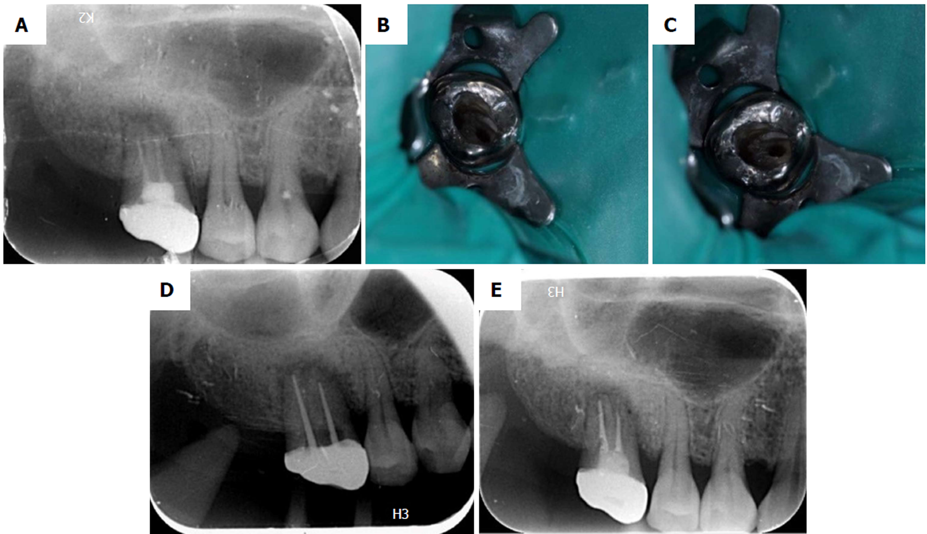
Figure 3 Treatment process using a dental operating microscope and X-ray images (Case 2).
A: Radiograph shows incomplete obturation of two root canals and the existence of a periapical lesion; B-C: Only two separate root canals are detected under the visual field of the dental operating microscope; D: Radiograph of master cone; E: Post-obturation periapical radiograph.
- Citation: Liu J, Que KH, Xiao ZH, Wen W. Endodontic management of the maxillary first molars with two root canals: A case report and review of the literature. World J Clin Cases 2019; 7(1): 79-88
- URL: https://www.wjgnet.com/2307-8960/full/v7/i1/79.htm
- DOI: https://dx.doi.org/10.12998/wjcc.v7.i1.79


