Copyright
©The Author(s) 2019.
World J Clin Cases. Jan 6, 2019; 7(1): 109-115
Published online Jan 6, 2019. doi: 10.12998/wjcc.v7.i1.109
Published online Jan 6, 2019. doi: 10.12998/wjcc.v7.i1.109
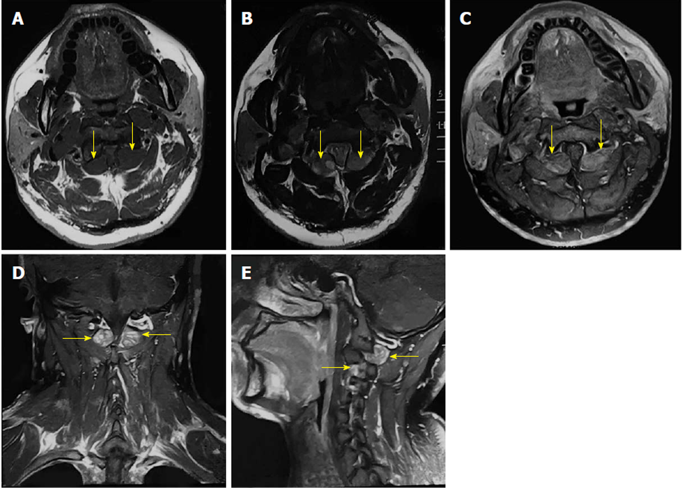
Figure 1 Preoperative magnetic resonance images of the patient.
A: Axial T1-weighted image shows bilateral dumbbell masses displaying low signal (arrows); B and D: Axial and coronal T2-weighted images show bilateral dumbbell masses displaying high signal (arrows); C and E: Axial and sagittal T1-weighted images after administration of gadolinium-diethylenetriaminepentaacetic acid depict prominent heterogeneous enhancement of the lesions (contrast dose: 15 mL: 7.04 g) (arrows).
- Citation: Tan CY, Liu JW, Lin Y, Tie XX, Cheng P, Qi X, Gao Y, Guo ZZ. Bilateral and symmetric C1-C2 dumbbell ganglioneuromas associated with neurofibromatosis type 1: A case report. World J Clin Cases 2019; 7(1): 109-115
- URL: https://www.wjgnet.com/2307-8960/full/v7/i1/109.htm
- DOI: https://dx.doi.org/10.12998/wjcc.v7.i1.109


