Copyright
©The Author(s) 2018.
World J Clin Cases. Sep 6, 2018; 6(9): 301-307
Published online Sep 6, 2018. doi: 10.12998/wjcc.v6.i9.301
Published online Sep 6, 2018. doi: 10.12998/wjcc.v6.i9.301
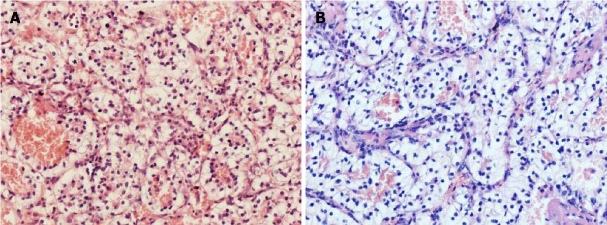
Figure 3 Histological findings.
A: H and E stain of the clival lesion, 200 ×; B: H and E stain of the left renal mass, 200 × showing clear cells with alveolar growth and separated by reticular separation of thin wall vessels. The two lesions demonstrated similar histopathologic features.
- Citation: Zhang WQ, Bao Y, Qiu B, Wang Y, Li ZP, Wang YB. Clival metastasis of renal clear cell carcinoma: Case report and literature review. World J Clin Cases 2018; 6(9): 301-307
- URL: https://www.wjgnet.com/2307-8960/full/v6/i9/301.htm
- DOI: https://dx.doi.org/10.12998/wjcc.v6.i9.301


