Copyright
©The Author(s) 2018.
World J Clin Cases. Jun 16, 2018; 6(6): 127-131
Published online Jun 16, 2018. doi: 10.12998/wjcc.v6.i6.127
Published online Jun 16, 2018. doi: 10.12998/wjcc.v6.i6.127
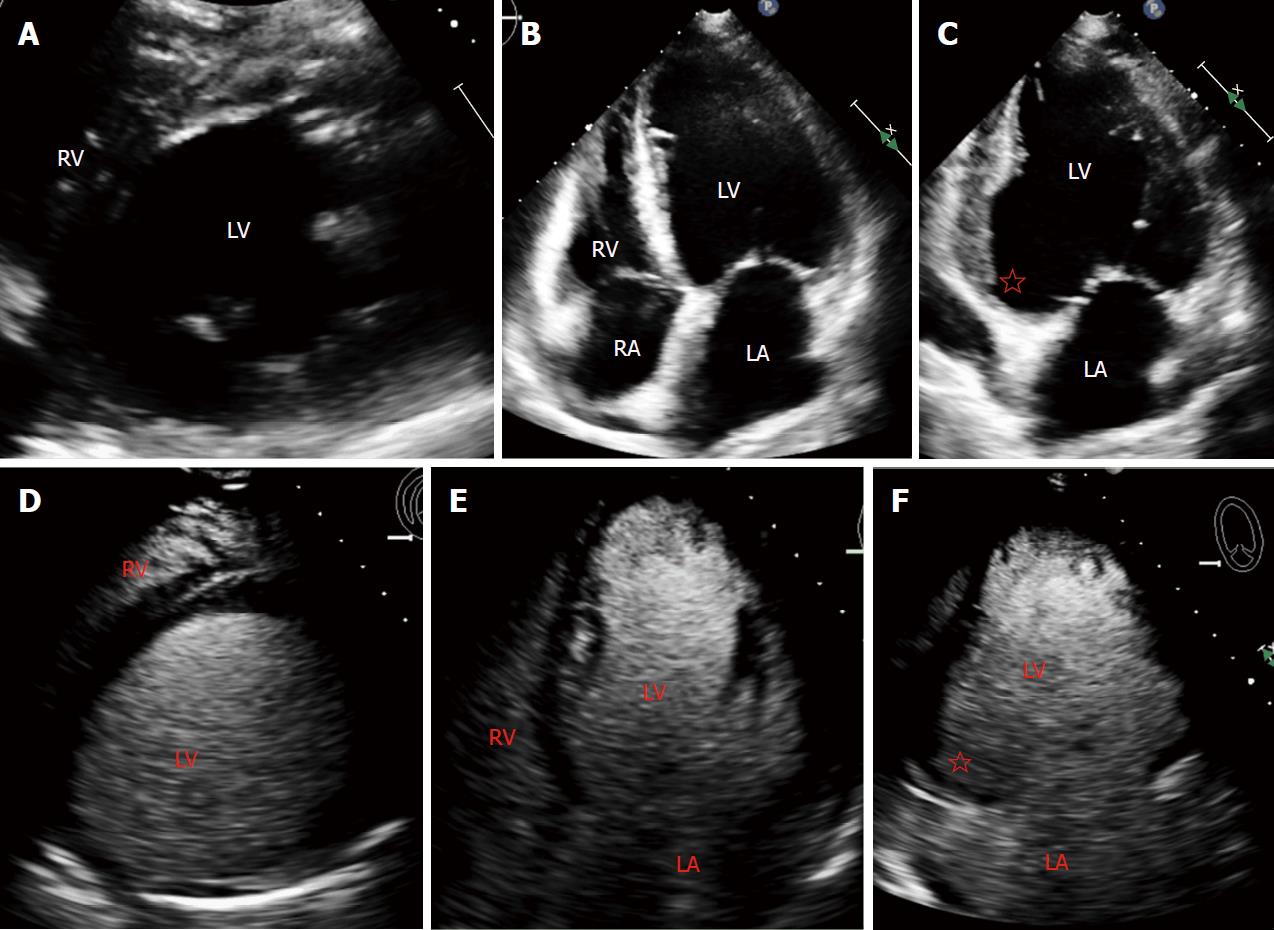
Figure 1 Transthoracic echocardiogram without and with definity contrast demonstrated absence of thrombus.
A: Parasternal short axis of transthoracic echocardiogram; B: Apical 4-chamber of transthoracic echocardiogram; C: Apical 2-chamber of transthoracic echocardiogram; D: Parasternal short axis transthoracic echocardiogram with definity contrast; E: Apical 4-chamber transthoracic echocardiogram with definity contrast; F: Apical 2-chamber transthoracic echocardiogram with definity contrast. There is an inferior wall aneurysm (red star). LA: Left atrium; LV: Left ventricle; RV: Right ventricle; RA: Right atrium.
- Citation: Siddiqui I, Nguyen T, Movahed A, Kabirdas D. Elusive left ventricular thrombus: Diagnostic role of cardiac magnetic resonance imaging-A case report and review of the literature. World J Clin Cases 2018; 6(6): 127-131
- URL: https://www.wjgnet.com/2307-8960/full/v6/i6/127.htm
- DOI: https://dx.doi.org/10.12998/wjcc.v6.i6.127


