Copyright
©The Author(s) 2018.
World J Clin Cases. May 16, 2018; 6(5): 88-93
Published online May 16, 2018. doi: 10.12998/wjcc.v6.i5.88
Published online May 16, 2018. doi: 10.12998/wjcc.v6.i5.88
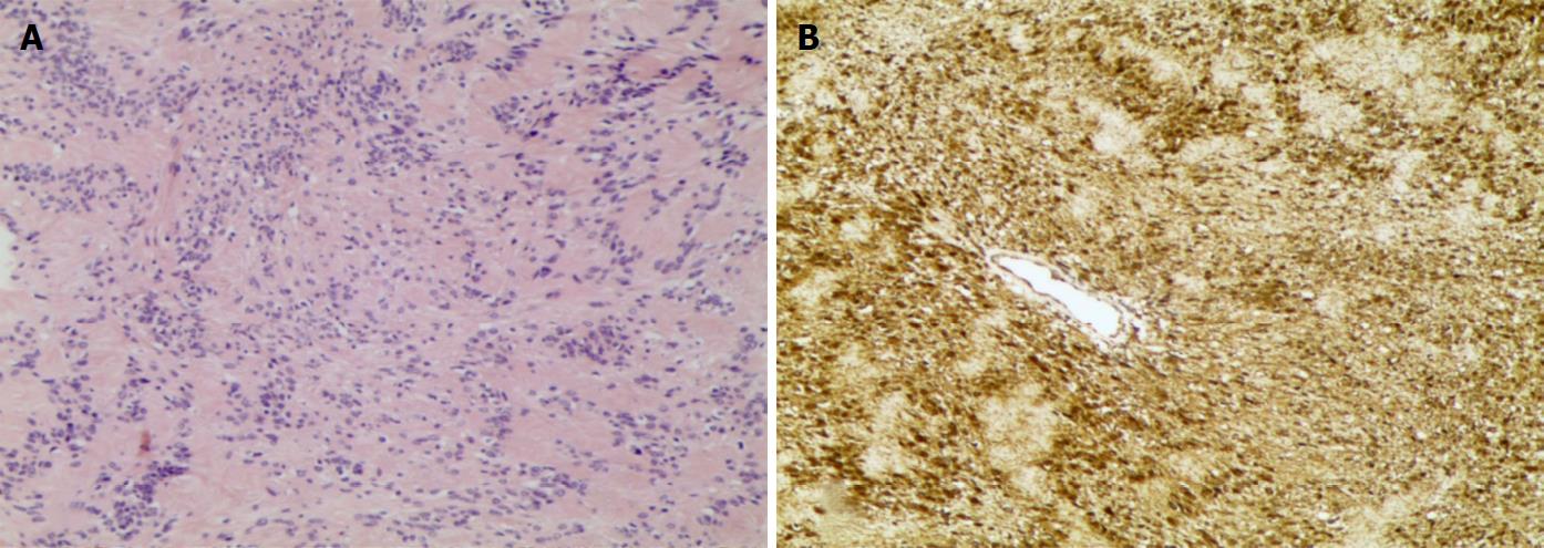
Figure 3 Pathological results.
A: Predominant Antoni A areas composed of spindle cells with palisading parallel rows are displayed by haematoxylin and eosin stain; B: Immunostain shows the tumour cells with diffuse immunoreactivity to S100 protein.
- Citation: Sun XL, Wen K, Xu ZZ, Wang XP. Magnetic resonance imaging findings for differential diagnosis of perianal plexiform schwannoma: Case report and review of the literature. World J Clin Cases 2018; 6(5): 88-93
- URL: https://www.wjgnet.com/2307-8960/full/v6/i5/88.htm
- DOI: https://dx.doi.org/10.12998/wjcc.v6.i5.88


