Copyright
©The Author(s) 2018.
World J Clin Cases. May 16, 2018; 6(5): 88-93
Published online May 16, 2018. doi: 10.12998/wjcc.v6.i5.88
Published online May 16, 2018. doi: 10.12998/wjcc.v6.i5.88
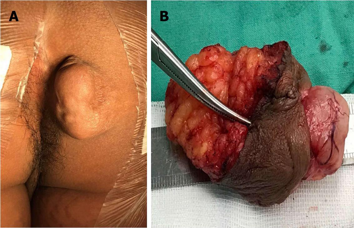
Figure 1 Macrography of plexiform schwannoma.
A: A multinodular cystic-solid mass of 5 cm × 3 cm is located on the right ischioanal fossa; B: The resected specimen shows a superficial vessel implanting into the tumour.
- Citation: Sun XL, Wen K, Xu ZZ, Wang XP. Magnetic resonance imaging findings for differential diagnosis of perianal plexiform schwannoma: Case report and review of the literature. World J Clin Cases 2018; 6(5): 88-93
- URL: https://www.wjgnet.com/2307-8960/full/v6/i5/88.htm
- DOI: https://dx.doi.org/10.12998/wjcc.v6.i5.88


