Copyright
©The Author(s) 2018.
World J Clin Cases. Feb 16, 2018; 6(2): 11-19
Published online Feb 16, 2018. doi: 10.12998/wjcc.v6.i2.11
Published online Feb 16, 2018. doi: 10.12998/wjcc.v6.i2.11
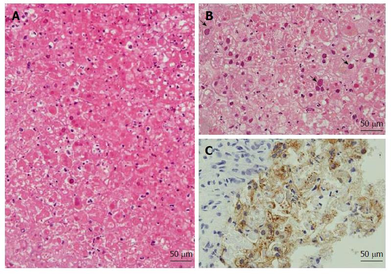
Figure 2 Histology of postmortem needle biopsies of the liver.
A: Massive hemorrhagic necrosis with minimum inflammatory infiltrates (hematoxylin-eosin stain; original magnification, 200 ×); B: Viral inclusions (arrow) scattered in infected hepatocytes (hematoxylin-eosin stain; original magnification, 200 ×); C: Immunostaining for herpes simplex virus type 1 (original magnification, 200 ×).
- Citation: Yokoi Y, Kaneko T, Sawayanagi T, Takano Y, Watahiki Y. Fatal fulminant herpes simplex hepatitis following surgery in an adult. World J Clin Cases 2018; 6(2): 11-19
- URL: https://www.wjgnet.com/2307-8960/full/v6/i2/11.htm
- DOI: https://dx.doi.org/10.12998/wjcc.v6.i2.11


