Copyright
©The Author(s) 2018.
World J Clin Cases. Dec 26, 2018; 6(16): 1217-1222
Published online Dec 26, 2018. doi: 10.12998/wjcc.v6.i16.1217
Published online Dec 26, 2018. doi: 10.12998/wjcc.v6.i16.1217
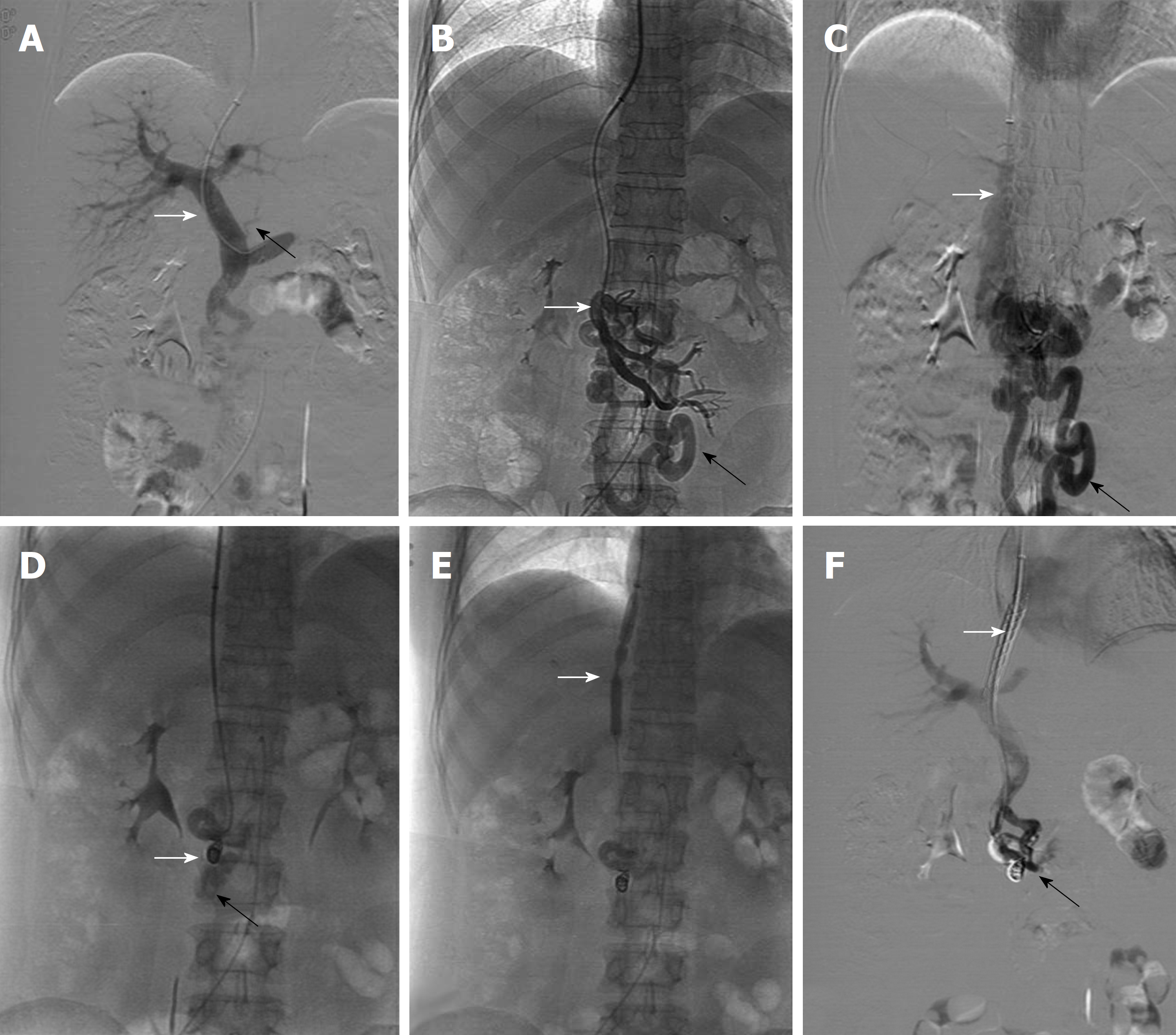
Figure 2 Imaging features of the transjugular intra-hepatic porto-systemic shunt procedure.
A: Transhepatic portogram using iodinated contrast material showed dilated main portal vein (white arrow) and mild gastric coronary vein (black arrow) after successful hepatic vein puncture to the port vein; B: Mesenteric angiography indicated the remarkably dilated duodenal varices (black arrow) extending from the proximal superior mesenteric vein (white arrow); C: The shunt (black arrow) extends from the superior mesenteric vein towards the inferior vena cava (white arrow); D: Mesenteric angiography illustrates that the dilated duodenal varices were obliterated by stainless-steel coils and Histoacryl. The deployed coils (white arrow) and reduced duodenal varices (black arrow) are clearly denoted in the fluoroscopic image; E: Balloon dilatation (white arrow) of a tract interposed between the hepatic and portal veins is shown in the fluoroscopic image; F: Portal venogram after transjugular intra-hepatic porto-systemic shunt insertion with a metallic bare stent and a covered stent (white arrow) demonstrates that the indicated iodinated contrast flows easily towards the heart. Peripheral portal vein branches are reduced because of flow reversal. The deployed coils and reduced duodenal varices (black arrow) are shown by portography.
- Citation: Xie BS, Zhong JW, Wang AJ, Zhang ZD, Zhu X, Guo GH. Duodenal variceal bleeding secondary to idiopathic portal hypertension treated with transjugular intra-hepatic porto-systemic shunt plus embolization: A case report. World J Clin Cases 2018; 6(16): 1217-1222
- URL: https://www.wjgnet.com/2307-8960/full/v6/i16/1217.htm
- DOI: https://dx.doi.org/10.12998/wjcc.v6.i16.1217


