Copyright
©The Author(s) 2018.
World J Clin Cases. Dec 26, 2018; 6(16): 1217-1222
Published online Dec 26, 2018. doi: 10.12998/wjcc.v6.i16.1217
Published online Dec 26, 2018. doi: 10.12998/wjcc.v6.i16.1217
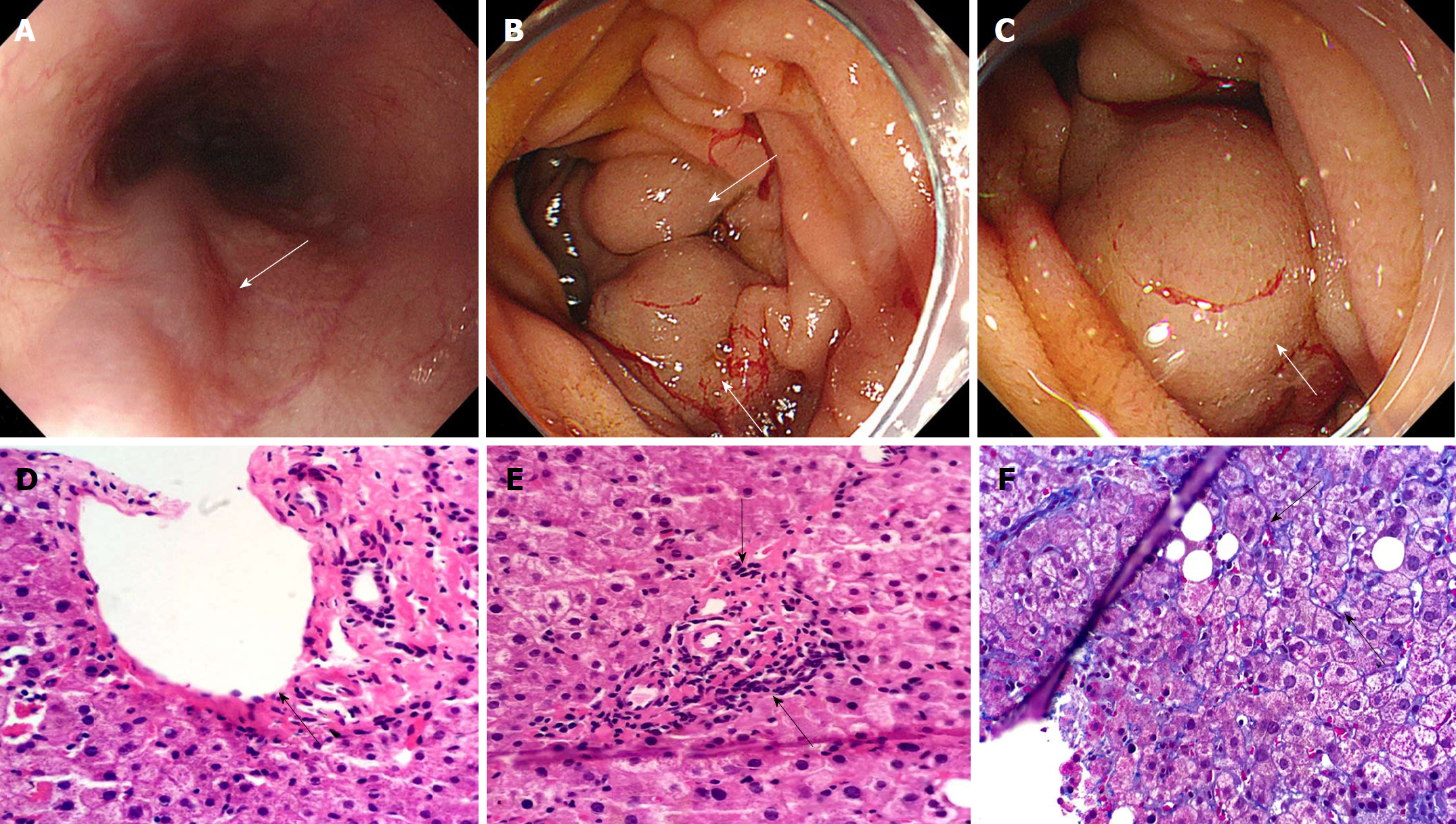
Figure 1 Imaging features of endoscopy and hepatic pathology.
A: An esophagogastroduodenoscopy (EGD) showed mild varices (white arrow) located in the esophagus without bloody scab; B and C: An EGD displayed a large submucosal vermicular mass (white arrow) located in the first and second duodenum with bloody scab; D and E: Liver samples were observed pathologically with Hematoxylin and Eosin staining. There was an integrated structure in the hepatic lobule with small sporadic necrotic foci, in which hepatic cells were moderately swollen with uneven nuclear size and fibrosis in portal triads. The extreme extension (D) and occlusion (E) of portal branches in portal regions are denoted by the black arrow; F: Perisinusoidal fibrosis (black arrow) was surrounded by hepatic lobule, as shown by Masson staining.
- Citation: Xie BS, Zhong JW, Wang AJ, Zhang ZD, Zhu X, Guo GH. Duodenal variceal bleeding secondary to idiopathic portal hypertension treated with transjugular intra-hepatic porto-systemic shunt plus embolization: A case report. World J Clin Cases 2018; 6(16): 1217-1222
- URL: https://www.wjgnet.com/2307-8960/full/v6/i16/1217.htm
- DOI: https://dx.doi.org/10.12998/wjcc.v6.i16.1217


