Copyright
©The Author(s) 2018.
World J Clin Cases. Dec 26, 2018; 6(16): 1210-1216
Published online Dec 26, 2018. doi: 10.12998/wjcc.v6.i16.1210
Published online Dec 26, 2018. doi: 10.12998/wjcc.v6.i16.1210
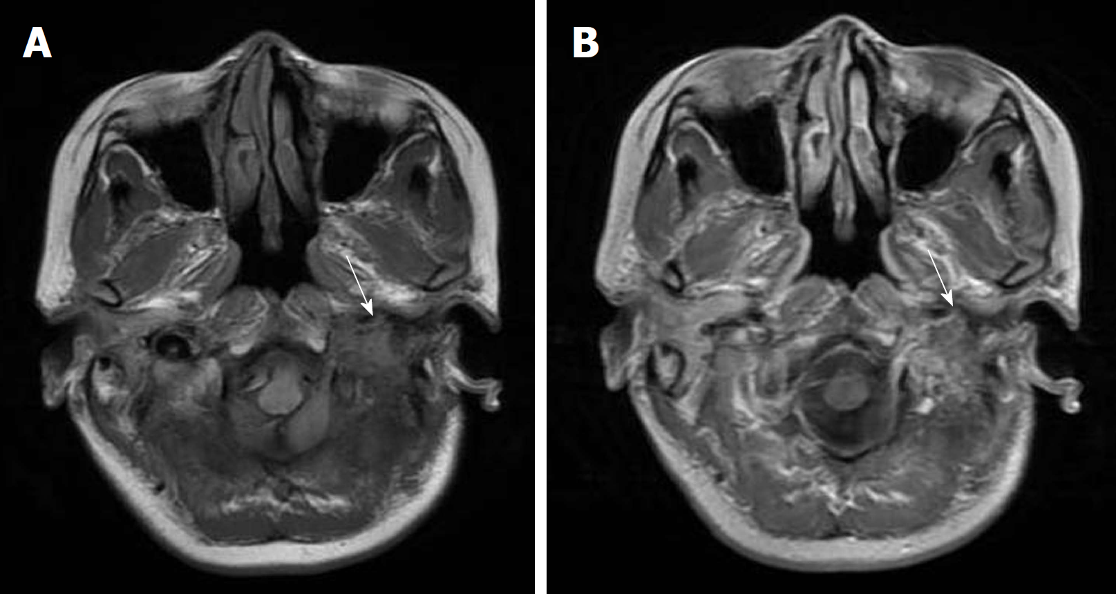
Figure 4 Postoperative magnetic resonance imaging scans.
A, B: The axial T1-weighted image (A) and gadolinium-enhanced T1-weighted image (B) show that the mass originated from the mastoid portion of the temporal bone and displayed contrast enhancement before it was excised (arrow).
- Citation: Zheng YM, Wang HX, Dong C. Chondromyxoid fibroma of the temporal bone: A case report and review of the literature. World J Clin Cases 2018; 6(16): 1210-1216
- URL: https://www.wjgnet.com/2307-8960/full/v6/i16/1210.htm
- DOI: https://dx.doi.org/10.12998/wjcc.v6.i16.1210


