Copyright
©The Author(s) 2018.
World J Clin Cases. Dec 26, 2018; 6(16): 1210-1216
Published online Dec 26, 2018. doi: 10.12998/wjcc.v6.i16.1210
Published online Dec 26, 2018. doi: 10.12998/wjcc.v6.i16.1210
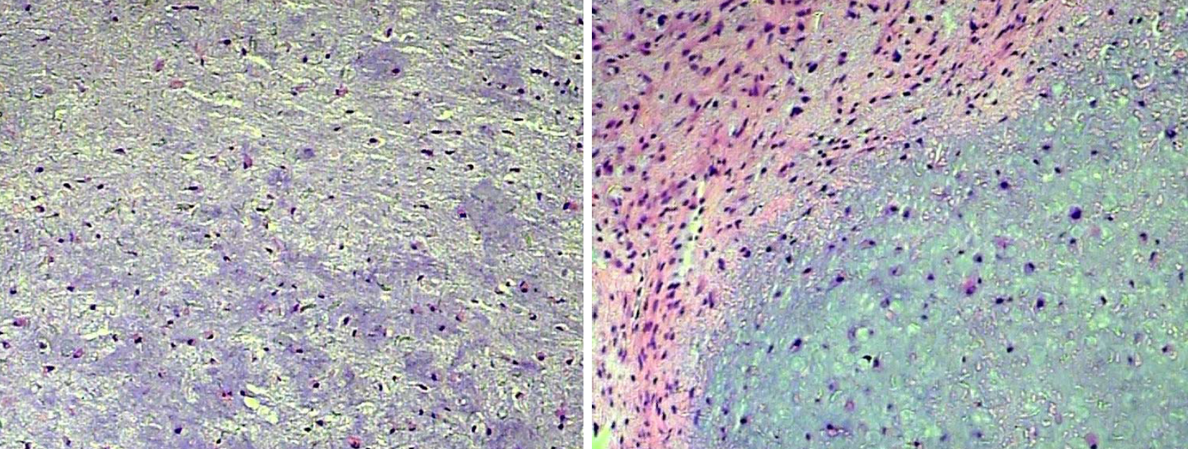
Figure 3 High-power view of resected specimen showing a myxoid lesion consisting of cartilage material, admixed with spindle-shaped cells and bland stromal cells (H and E staining, ×200).
- Citation: Zheng YM, Wang HX, Dong C. Chondromyxoid fibroma of the temporal bone: A case report and review of the literature. World J Clin Cases 2018; 6(16): 1210-1216
- URL: https://www.wjgnet.com/2307-8960/full/v6/i16/1210.htm
- DOI: https://dx.doi.org/10.12998/wjcc.v6.i16.1210


