Copyright
©The Author(s) 2018.
World J Clin Cases. Dec 26, 2018; 6(16): 1210-1216
Published online Dec 26, 2018. doi: 10.12998/wjcc.v6.i16.1210
Published online Dec 26, 2018. doi: 10.12998/wjcc.v6.i16.1210
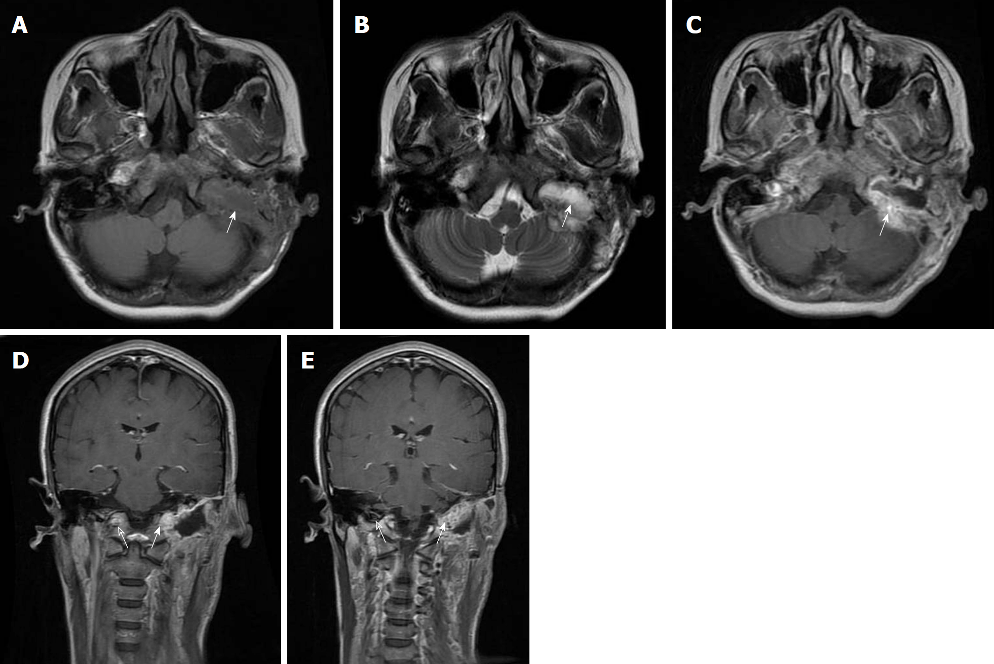
Figure 2 Magnetic resonance imaging findings of the lesion.
A-C: The lesion is hypointensity on axial T1-weighted image (A, arrow), heterogeneous hyperintensity on axial T2-weighted image (B, arrow), and enhances peripherally (C, arrow); D, E: The coronal contrast enhanced MR images show that the tumour invaded the left hypoglossal canal (D, arrow) and the left jugular foramen (E, arrow). The contralateral hypoglossal canal (D, blank arrow) and the jugular foramen (E, blank arrow) were normal.
- Citation: Zheng YM, Wang HX, Dong C. Chondromyxoid fibroma of the temporal bone: A case report and review of the literature. World J Clin Cases 2018; 6(16): 1210-1216
- URL: https://www.wjgnet.com/2307-8960/full/v6/i16/1210.htm
- DOI: https://dx.doi.org/10.12998/wjcc.v6.i16.1210


