Copyright
©The Author(s) 2018.
World J Clin Cases. Dec 26, 2018; 6(16): 1182-1188
Published online Dec 26, 2018. doi: 10.12998/wjcc.v6.i16.1182
Published online Dec 26, 2018. doi: 10.12998/wjcc.v6.i16.1182
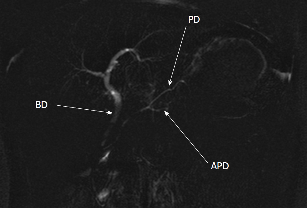
Figure 3 Magnetic resonance cholangiopancreatography shows the pancreatic duplication.
The accessory pancreatic duct of the accessory pancreatic lobe is located ventrally to the main pancreatic duct. The confluence is located in the body of the pancreas. The biliary duct is of common appearance. Published with permission of Jablonec nad Nisou Hospital. APD: Accessory pancreatic duct; PD: Pancreatic duct; BD: Biliary duct.
- Citation: Rousek M, Kachlik D, Nikov A, Pintova J, Ryska M. Gastric duplication cyst communicating to accessory pancreatic lobe: A case report and review of the literature. World J Clin Cases 2018; 6(16): 1182-1188
- URL: https://www.wjgnet.com/2307-8960/full/v6/i16/1182.htm
- DOI: https://dx.doi.org/10.12998/wjcc.v6.i16.1182


