Copyright
©The Author(s) 2018.
World J Clin Cases. Dec 26, 2018; 6(16): 1175-1181
Published online Dec 26, 2018. doi: 10.12998/wjcc.v6.i16.1175
Published online Dec 26, 2018. doi: 10.12998/wjcc.v6.i16.1175
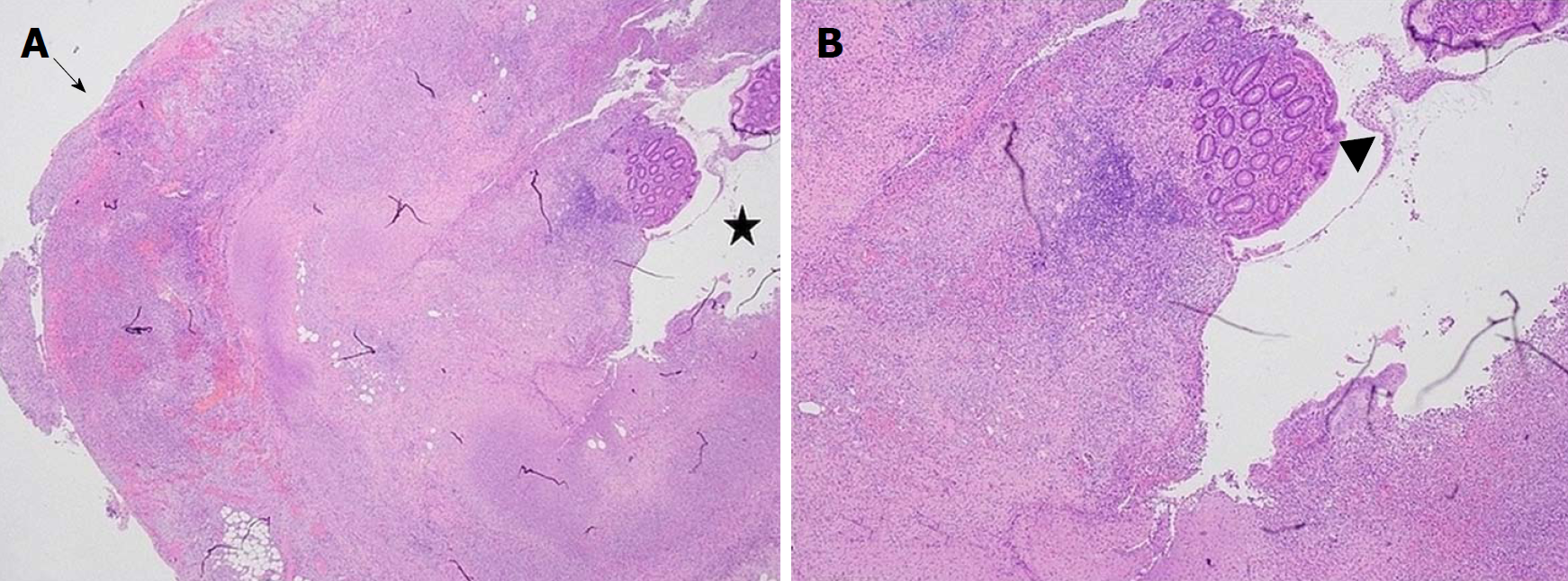
Figure 1 Histopathology of appendiceal specimen with appendicitis.
A: This slide is a cross-section of the appendix showing the appendiceal lumen indicated by the star displaying extensive ulceration and pervasive inflammation; with minimal residual mucosa. The transmural inflammation, involves the serosa as shown by the arrow; and depicts mucosal ulceration and necrosis; B: This slide image, photographed at a higher magnification, shows the residual mucosa (which appears normal) surrounded by necrotic tissue and cellular debris. The residual mucosa is indicated by the arrowhead.
- Citation: Tse A, Cheluvappa R, Selvendran S. Post-appendectomy pelvic abscess with extended-spectrum beta-lactamase producing Escherichia coli: A case report and review of literature. World J Clin Cases 2018; 6(16): 1175-1181
- URL: https://www.wjgnet.com/2307-8960/full/v6/i16/1175.htm
- DOI: https://dx.doi.org/10.12998/wjcc.v6.i16.1175


