Copyright
©The Author(s) 2018.
World J Clin Cases. Dec 26, 2018; 6(16): 1164-1168
Published online Dec 26, 2018. doi: 10.12998/wjcc.v6.i16.1164
Published online Dec 26, 2018. doi: 10.12998/wjcc.v6.i16.1164
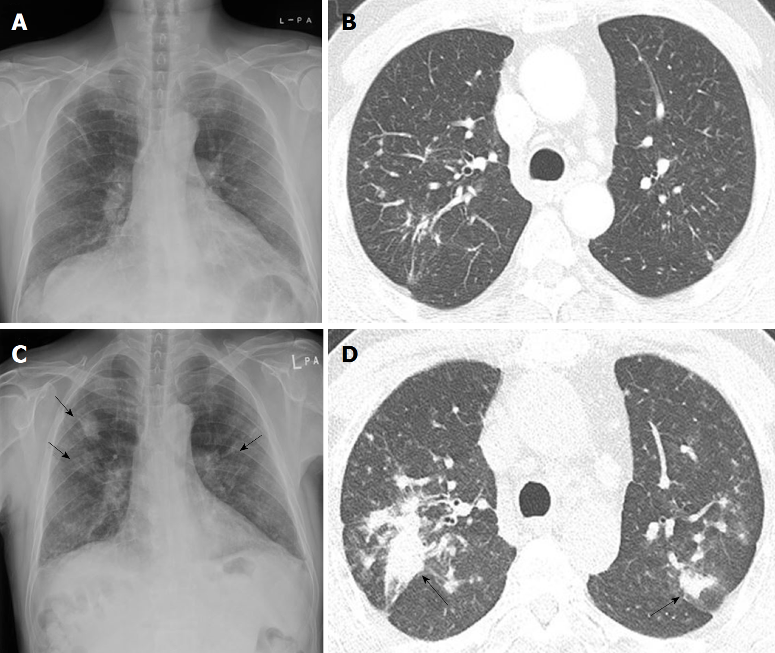
Figure 1 Serial chest radiographs and computed tomography scans of a 64-year-old man with rapidly progressing pneumoconiosis.
A: Radiograph in November 2011 shows poorly-defined small nodules and fibrotic linear opacities in both upper lungs; B: Multiple poorly-defined small nodules were observed in both upper lobes, predominantly in posterior segments, on initial computed tomography (CT) scan obtained in November 2011. Minimal fibrotic linear opacities were also noted; C: Follow-up radiograph in February 2018 demonstrated significant increase in multiple small nodules in both lungs, with new poorly-defined large nodules in both upper lungs (arrows); D: Follow-up CT scan in February 2018 showed increased size and number of small poorly-defined nodules in both upper lobes, with ill-defined mass formation in posterior segments (arrows).
- Citation: Yoon HY, Kim Y, Park HS, Kang CW, Ryu YJ. Combined silicosis and mixed dust pneumoconiosis with rapid progression: A case report and literature review. World J Clin Cases 2018; 6(16): 1164-1168
- URL: https://www.wjgnet.com/2307-8960/full/v6/i16/1164.htm
- DOI: https://dx.doi.org/10.12998/wjcc.v6.i16.1164


