Copyright
©The Author(s) 2018.
World J Clin Cases. Dec 6, 2018; 6(15): 1059-1066
Published online Dec 6, 2018. doi: 10.12998/wjcc.v6.i15.1059
Published online Dec 6, 2018. doi: 10.12998/wjcc.v6.i15.1059
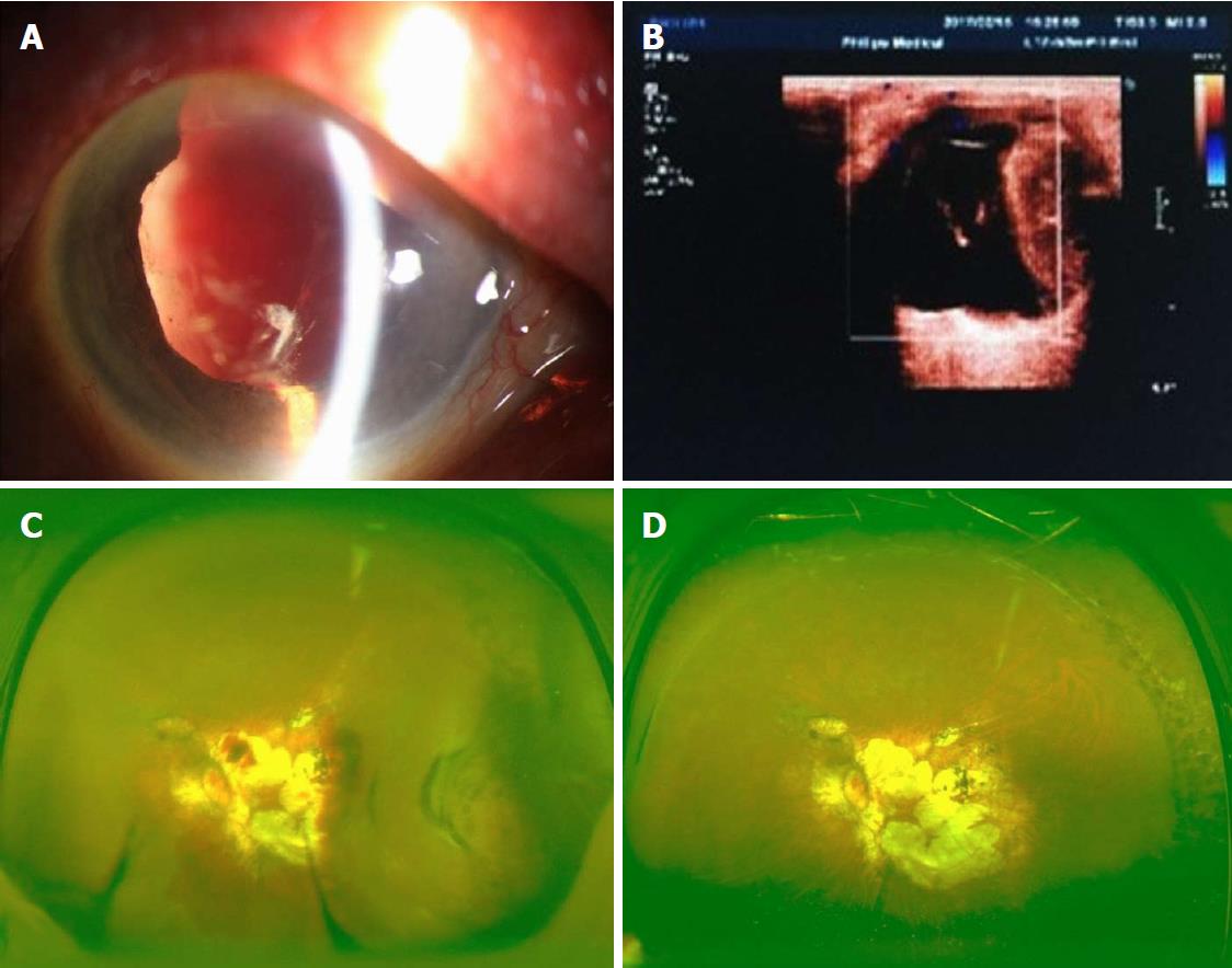
Figure 2 Clinical findings in patient 2.
A: An examination showed a deep anterior chamber with blood and cells, iridocoloboma, aphakia, capsule remnants, and a massive vitreous hemorrhage; B: A color ultrasound showed choroidal detachment; C: The day after PPV, a globular elevation of the choroidal detachment was clearly visible in the temporal quadrant; D: At 1 year later, the fundus was flat without any signs of choroidal detachment.
- Citation: Chai F, Ai H, Deng J, Zhao XQ. Sub-Tenon’s urokinase injection-assisted vitrectomy in early treatment of suprachoroidal hemorrhage: Four cases report. World J Clin Cases 2018; 6(15): 1059-1066
- URL: https://www.wjgnet.com/2307-8960/full/v6/i15/1059.htm
- DOI: https://dx.doi.org/10.12998/wjcc.v6.i15.1059


