Copyright
©The Author(s) 2018.
World J Clin Cases. Dec 6, 2018; 6(15): 1059-1066
Published online Dec 6, 2018. doi: 10.12998/wjcc.v6.i15.1059
Published online Dec 6, 2018. doi: 10.12998/wjcc.v6.i15.1059
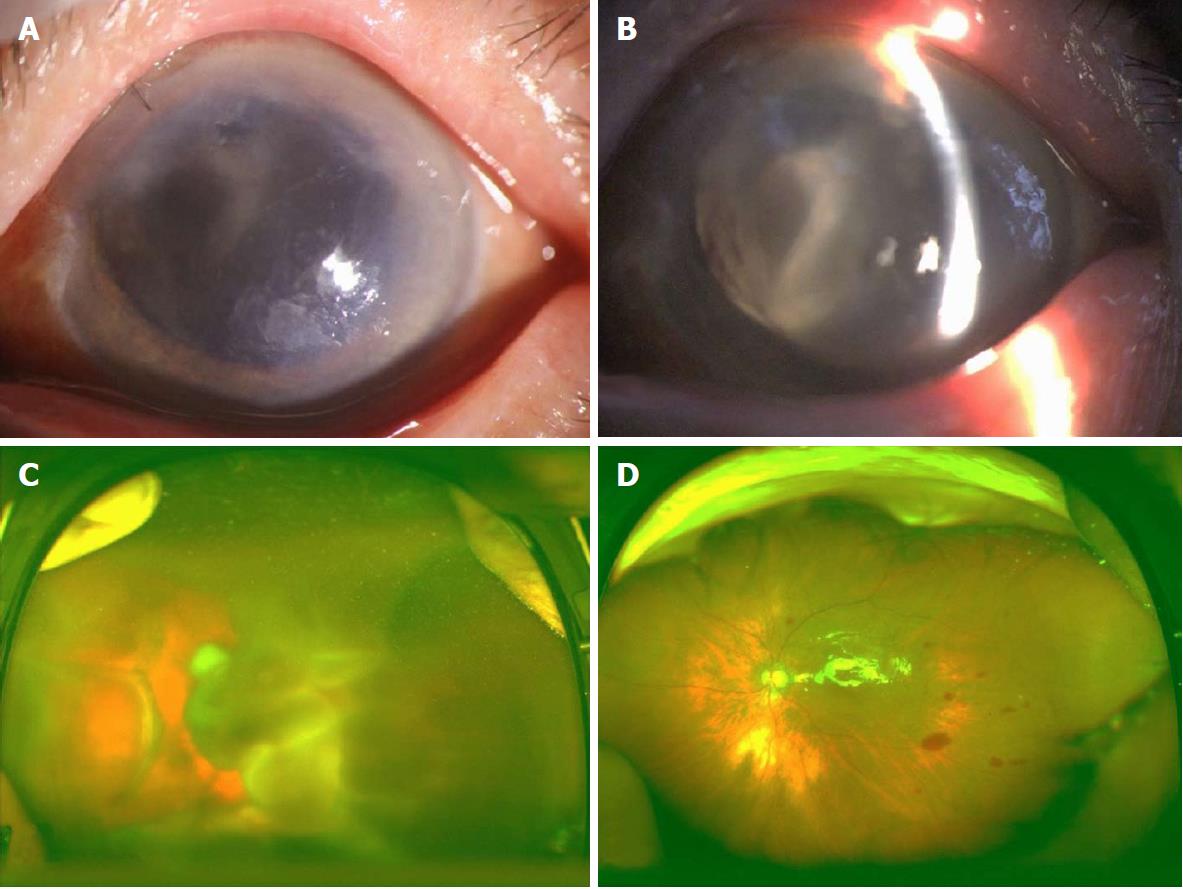
Figure 1 Clinical findings in patient 1.
A: The cornea exhibited edema with local epithelial defect; B: The left eye was aphakic, and a prominent retinal detachment was visible through the pupil; C: A fundus examination revealed retinal detachment with choroidal detachment; D: Postoperatively, the retinal and choroidal detachment was completely reduced.
- Citation: Chai F, Ai H, Deng J, Zhao XQ. Sub-Tenon’s urokinase injection-assisted vitrectomy in early treatment of suprachoroidal hemorrhage: Four cases report. World J Clin Cases 2018; 6(15): 1059-1066
- URL: https://www.wjgnet.com/2307-8960/full/v6/i15/1059.htm
- DOI: https://dx.doi.org/10.12998/wjcc.v6.i15.1059


