Copyright
©The Author(s) 2018.
World J Clin Cases. Dec 6, 2018; 6(15): 1036-1041
Published online Dec 6, 2018. doi: 10.12998/wjcc.v6.i15.1036
Published online Dec 6, 2018. doi: 10.12998/wjcc.v6.i15.1036
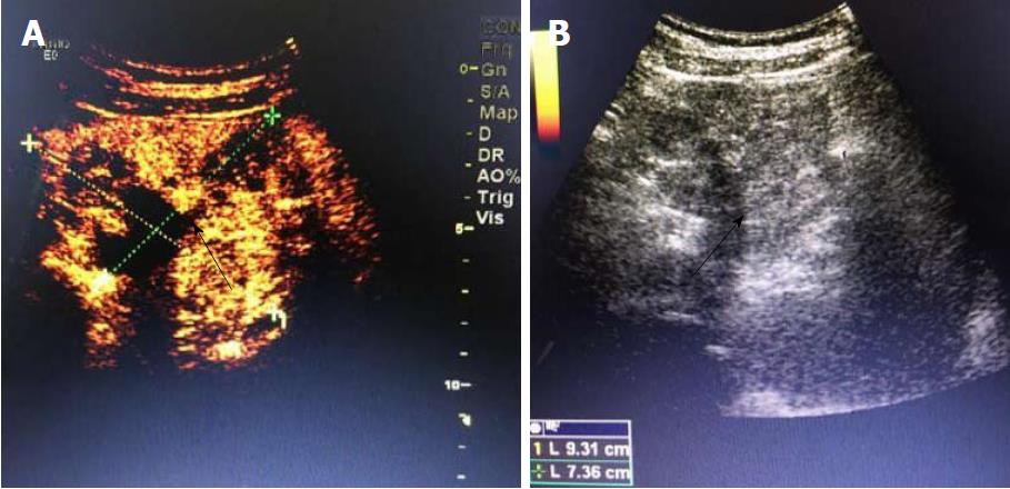
Figure 4 Findings of abdominal contrast-enhanced ultrasonography.
A: Abdominal contrast-enhanced ultrasonography shows an abnormal morphology of the pancreas. A mass with inhomogeneous echo, sized about 9.3 cm × 7.2 cm, is found in the anterior pancreatic body; it has a well-defined border and regular shape, but the border with the parenchyma of pancreatic body and tail is poorly defined, with flocculent echo and strong spotty or patchy echo in the central part; B: Color Doppler flow imaging showed a few low-speed arteriovenous blood flow signals, whereas the echoes are homogeneous in the remaining parenchyma. The main pancreatic duct is not dilated. Black arrows indicate the mass.
- Citation: Zhang MY, Tian BL. Pancreatic panniculitis and solid pseudopapillary tumor of the pancreas: A case report. World J Clin Cases 2018; 6(15): 1036-1041
- URL: https://www.wjgnet.com/2307-8960/full/v6/i15/1036.htm
- DOI: https://dx.doi.org/10.12998/wjcc.v6.i15.1036


