Copyright
©The Author(s) 2018.
World J Clin Cases. Dec 6, 2018; 6(15): 1036-1041
Published online Dec 6, 2018. doi: 10.12998/wjcc.v6.i15.1036
Published online Dec 6, 2018. doi: 10.12998/wjcc.v6.i15.1036
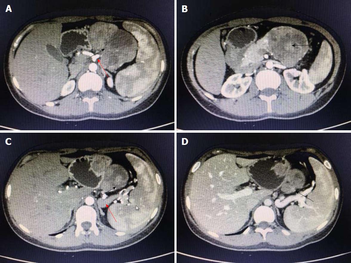
Figure 3 Findings of contrast-enhanced abdominal computed tomography scan.
A and B: Contrast-enhanced abdominal computed tomography reveals a mixed density mass with solid and cystic components, sized 6.8 cm × 7.8 cm × 8.8 cm, in the body and tail of the pancreas, with a well-defined border. Nodular calcifications are found inside the lesion; C: The solid component is obviously enhanced, although the degree of enhancement is lower than the normal pancreatic parenchyma; D: The spleen is slightly larger, and the splenic vein is compressed. Multiple collateral circulations are seen in the left upper abdomen. The lesion is fed by the common hepatic artery and a branch of splenic artery (red arrows in A and C, black arrows indicate the mass).
- Citation: Zhang MY, Tian BL. Pancreatic panniculitis and solid pseudopapillary tumor of the pancreas: A case report. World J Clin Cases 2018; 6(15): 1036-1041
- URL: https://www.wjgnet.com/2307-8960/full/v6/i15/1036.htm
- DOI: https://dx.doi.org/10.12998/wjcc.v6.i15.1036


