Copyright
©The Author(s) 2018.
World J Clin Cases. Dec 6, 2018; 6(15): 1012-1017
Published online Dec 6, 2018. doi: 10.12998/wjcc.v6.i15.1012
Published online Dec 6, 2018. doi: 10.12998/wjcc.v6.i15.1012
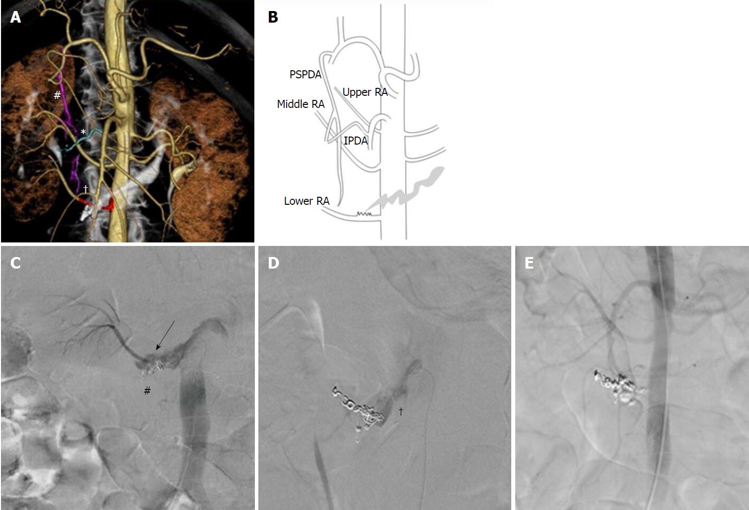
Figure 3 Embolization of the lower branch of the right renal artery.
A: 3D volume-rendered computed tomography image obtained during aortography. #Posterior superior pancreaticoduodenal artery (PSPDA; pink); *Inferior pancreaticoduodenal artery (IPDA; blue); †Lower branch of the right renal artery (lower RA; red); B: Schematic of the computed tomography image; C: Extravasation of contrast material is evident before embolization (arrow). #Endoscopic clips; D: Right renal arteriography after coil embolization shows residual extravasation (†); E: Aortography after embolization using coils and n-butyl-2-cyanoacrylate glue shows no extravasation.
- Citation: Anami S, Minamiguchi H, Shibata N, Koyama T, Sato H, Ikoma A, Nakai M, Yamagami T, Sonomura T. Successful endovascular treatment of endoscopically unmanageable hemorrhage from a duodenal ulcer fed by a renal artery: A case report. World J Clin Cases 2018; 6(15): 1012-1017
- URL: https://www.wjgnet.com/2307-8960/full/v6/i15/1012.htm
- DOI: https://dx.doi.org/10.12998/wjcc.v6.i15.1012


