Copyright
©The Author(s) 2018.
World J Clin Cases. Nov 26, 2018; 6(14): 854-861
Published online Nov 26, 2018. doi: 10.12998/wjcc.v6.i14.854
Published online Nov 26, 2018. doi: 10.12998/wjcc.v6.i14.854
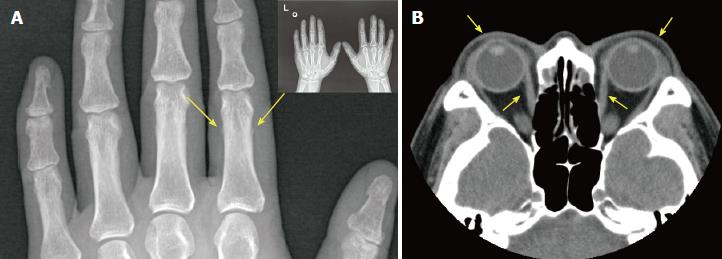
Figure 3 Imaging results of hands and eye socket.
A: X-ray scan showing the periosteal reaction in the phalangeal bone of the index finger of the left hand; B: Computed tomography scan of the eye socket showing that the bilateral extraocular muscles were slightly thickened and that both eyeballs extruded slightly.
- Citation: Zhang F, Lin XY, Chen J, Peng SQ, Shan ZY, Teng WP, Yu XH. Intralesional and topical glucocorticoids for pretibial myxedema: A case report and review of literature. World J Clin Cases 2018; 6(14): 854-861
- URL: https://www.wjgnet.com/2307-8960/full/v6/i14/854.htm
- DOI: https://dx.doi.org/10.12998/wjcc.v6.i14.854


