Copyright
©The Author(s) 2018.
World J Clin Cases. Nov 26, 2018; 6(14): 847-853
Published online Nov 26, 2018. doi: 10.12998/wjcc.v6.i14.847
Published online Nov 26, 2018. doi: 10.12998/wjcc.v6.i14.847
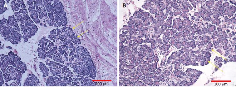
Figure 3 Histopathologic examination of the resected specimen.
Microscopic appearance showing that the lesion consisted of heterotopic pancreatic tissue (yellow arrows), including acini, islet cells, and pancreatic ducts, extending to the jejunal serosa (H and E staining; A: Magnification, × 100, scale bar = 200 μm; B: Magnification, × 200, scale bar = 100 μm).
- Citation: Tang XB, Liao MY, Wang WL, Bai YZ. Mesenteric heterotopic pancreas in a pediatric patient: A case report and review of literature. World J Clin Cases 2018; 6(14): 847-853
- URL: https://www.wjgnet.com/2307-8960/full/v6/i14/847.htm
- DOI: https://dx.doi.org/10.12998/wjcc.v6.i14.847


