Copyright
©The Author(s) 2018.
World J Clin Cases. Nov 26, 2018; 6(14): 847-853
Published online Nov 26, 2018. doi: 10.12998/wjcc.v6.i14.847
Published online Nov 26, 2018. doi: 10.12998/wjcc.v6.i14.847
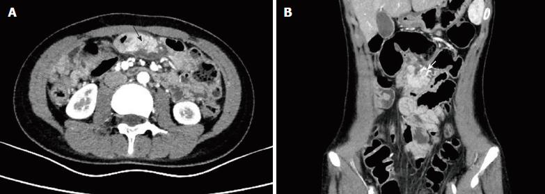
Figure 1 Contrast-enhanced computed tomography images of the abdomen.
A: Axial contrast-enhanced computed tomography (CECT) image of the abdomen showing an enhanced oval, soft-tissue mass in the jejunal mesentery at the level of the umbilicus (black arrow); B: Coronal CECT image showing that the mass had its own blood supply (white arrow).
- Citation: Tang XB, Liao MY, Wang WL, Bai YZ. Mesenteric heterotopic pancreas in a pediatric patient: A case report and review of literature. World J Clin Cases 2018; 6(14): 847-853
- URL: https://www.wjgnet.com/2307-8960/full/v6/i14/847.htm
- DOI: https://dx.doi.org/10.12998/wjcc.v6.i14.847


