Copyright
©The Author(s) 2018.
World J Clin Cases. Nov 26, 2018; 6(14): 830-835
Published online Nov 26, 2018. doi: 10.12998/wjcc.v6.i14.830
Published online Nov 26, 2018. doi: 10.12998/wjcc.v6.i14.830
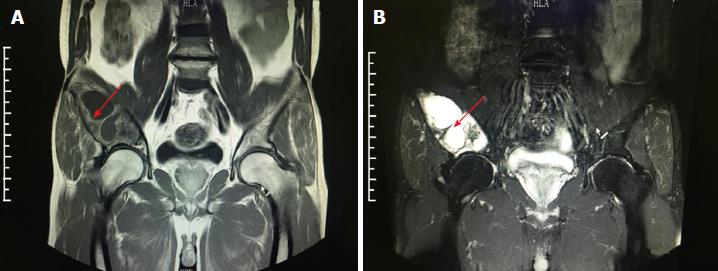
Figure 2 Magnetic resonance imaging examination results.
A: Extensive bone destruction in right ilium. T1w suggests low-density change; B: T2w suggests high-density change, with surrounding soft issue swelling (arrow indicates the area of lesion).
- Citation: Liu YB, Zou TM. Giant monostotic osteofibrous dysplasia of the ilium: A case report and review of literature. World J Clin Cases 2018; 6(14): 830-835
- URL: https://www.wjgnet.com/2307-8960/full/v6/i14/830.htm
- DOI: https://dx.doi.org/10.12998/wjcc.v6.i14.830


