Copyright
©The Author(s) 2018.
World J Clin Cases. Nov 26, 2018; 6(14): 820-824
Published online Nov 26, 2018. doi: 10.12998/wjcc.v6.i14.820
Published online Nov 26, 2018. doi: 10.12998/wjcc.v6.i14.820
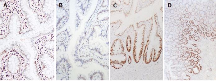
Figure 3 Immunohistochemichal staining for HES1, MLH1 and Ki67 in solitary rectal ulcer syndrome with sessile serrated adenomas/polyp.
A: Loss or very weak of nuclear expression of HES1 in crypts from sessile serrated adenomas/polyp (SSA/P) (Original magnification, × 100); B: Reduced number of surface epithelial cells expressing MLH1 protein in serrated crypts (Original magnification, × 100); C: Increased strong nuclear staining of Ki67 in the surface epithelium of solitary rectal ulcer syndrome with SSA/P architecture (Original magnification, × 100); D: Basal staining of Ki67 in glands of micro-vesicular hyperplastic polyps (Original magnification, × 40).
- Citation: Sun H, Sheng WQ, Huang D. Solitary rectal ulcer syndrome complicating sessile serrated adenoma/polyps: A case report and review of literature. World J Clin Cases 2018; 6(14): 820-824
- URL: https://www.wjgnet.com/2307-8960/full/v6/i14/820.htm
- DOI: https://dx.doi.org/10.12998/wjcc.v6.i14.820


