Copyright
©The Author(s) 2018.
World J Clin Cases. Nov 26, 2018; 6(14): 820-824
Published online Nov 26, 2018. doi: 10.12998/wjcc.v6.i14.820
Published online Nov 26, 2018. doi: 10.12998/wjcc.v6.i14.820
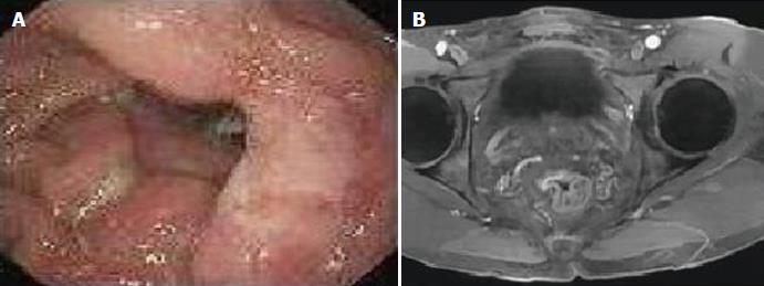
Figure 1 Endoscopic and magnetic resonance imaging of solitary rectal ulcer syndrome with sessile serrated adenomas/polyp.
A: Endoscopic imaging revealed a large ulcerative nodule in the anterior wall of the rectum; B: Magnetic resonance imagining showed focal thickening of the anterior rectal wall and irregularities in the mucosal surface.
- Citation: Sun H, Sheng WQ, Huang D. Solitary rectal ulcer syndrome complicating sessile serrated adenoma/polyps: A case report and review of literature. World J Clin Cases 2018; 6(14): 820-824
- URL: https://www.wjgnet.com/2307-8960/full/v6/i14/820.htm
- DOI: https://dx.doi.org/10.12998/wjcc.v6.i14.820


