Copyright
©The Author(s) 2018.
World J Clin Cases. Nov 26, 2018; 6(14): 800-806
Published online Nov 26, 2018. doi: 10.12998/wjcc.v6.i14.800
Published online Nov 26, 2018. doi: 10.12998/wjcc.v6.i14.800
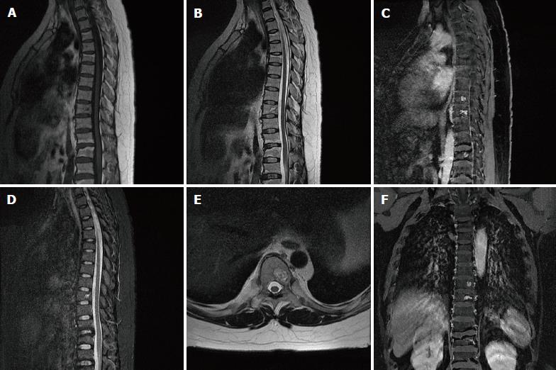
Figure 4 Spine magnetic resonance imaging of Patient 1.
A-F: Patchy bone lesions that were of low signal intensity on T1WI (A), mixed signal intensity on T2WI in sagittal (B), axial (E), and coronal images (F), and high signal intensity on fat saturated T2WI (C) with enhancement on gadolinium enhanced T1WI (D).
- Citation: Li S, Duan L, Wang FD, Lu L, Jin ZY. Carney complex: Two case reports and review of literature. World J Clin Cases 2018; 6(14): 800-806
- URL: https://www.wjgnet.com/2307-8960/full/v6/i14/800.htm
- DOI: https://dx.doi.org/10.12998/wjcc.v6.i14.800


