Copyright
©The Author(s) 2018.
World J Clin Cases. Nov 26, 2018; 6(14): 800-806
Published online Nov 26, 2018. doi: 10.12998/wjcc.v6.i14.800
Published online Nov 26, 2018. doi: 10.12998/wjcc.v6.i14.800
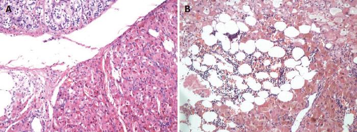
Figure 1 Histopathology (H and E staining, × 100).
A: The left adrenal lesion of Patient 1 conforms to primary pigmented nodular adrenocortical disease (PPNAD); B: The right adrenal lesion of Patient 2 conforms to PPNAD with local myelolipoma like change.
- Citation: Li S, Duan L, Wang FD, Lu L, Jin ZY. Carney complex: Two case reports and review of literature. World J Clin Cases 2018; 6(14): 800-806
- URL: https://www.wjgnet.com/2307-8960/full/v6/i14/800.htm
- DOI: https://dx.doi.org/10.12998/wjcc.v6.i14.800


