Copyright
©The Author(s) 2018.
World J Clin Cases. Nov 6, 2018; 6(13): 703-706
Published online Nov 6, 2018. doi: 10.12998/wjcc.v6.i13.703
Published online Nov 6, 2018. doi: 10.12998/wjcc.v6.i13.703
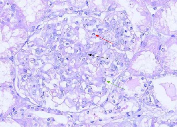
Figure 3 Light microscopy of the biopsied kidney tissue.
The mesangial area is moderately enlarged due to an increase in mesangial cells (black arrow) and the matrix. Endothelial cells (green arrow) show diffuse proliferation and degeneration. Infiltrated neutrophils (red arrow) are present [hematoxylin and eosin (HE) staining, × 200].
- Citation: Li XL, Ma ZG, Huang WH, Chai EQ, Hao YF. Successful treatment of pyoderma gangrenosum with concomitant immunoglobulin A nephropathy: A case report and review of literature. World J Clin Cases 2018; 6(13): 703-706
- URL: https://www.wjgnet.com/2307-8960/full/v6/i13/703.htm
- DOI: https://dx.doi.org/10.12998/wjcc.v6.i13.703


