Copyright
©The Author(s) 2018.
World J Clin Cases. Nov 6, 2018; 6(13): 683-687
Published online Nov 6, 2018. doi: 10.12998/wjcc.v6.i13.683
Published online Nov 6, 2018. doi: 10.12998/wjcc.v6.i13.683
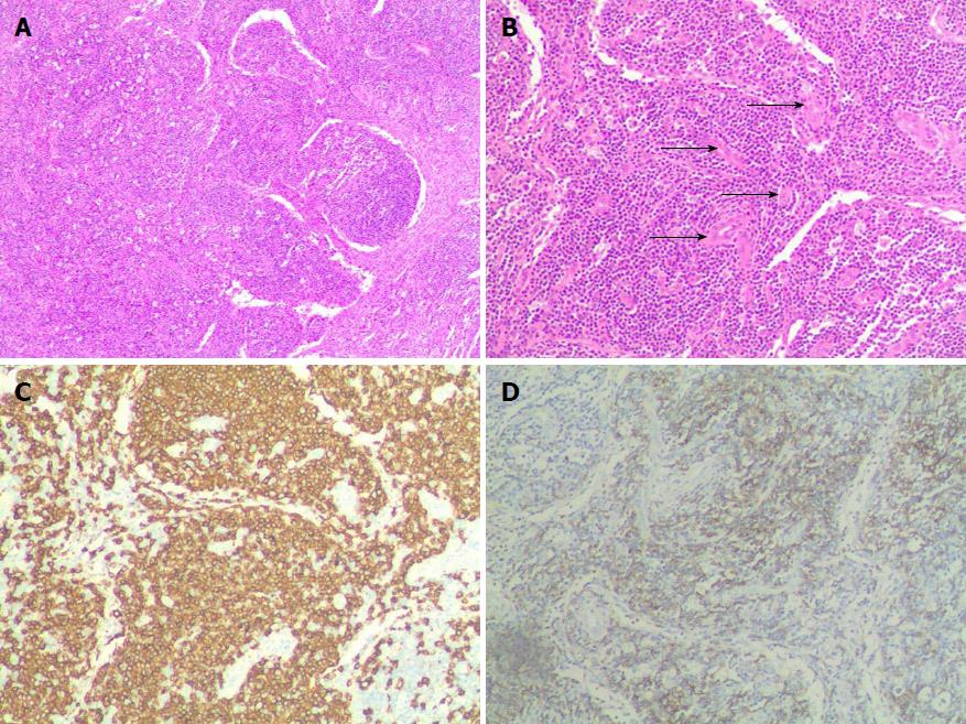
Figure 2 Pathological diagnosis of castleman disease was made by the analysis of morphological pattern and immunohistochemical markers.
A: hematoxylin-eosin (HE) (40 × magnification) staining showed a large number of enlarged lymphoid follicle scattered in the distribution; B: HE (100 × magnification) staining showed hyperplasia and wall thickening of the small blood vessels; C: Immunohistochemical staining (100 × magnification) showed CD20 was ubiquitously expressed; D: Immunohistochemical staining (HE 100 × magnification) showed CD21 was ubiquitously expressed. CD: Castleman disease; HE: Hematoxylin-eosin.
- Citation: Chen J, Yang C, Liang CZ. Detection of a unicentric type of Castleman-like mass at the site of adrenal grand: A case report and review of literature. World J Clin Cases 2018; 6(13): 683-687
- URL: https://www.wjgnet.com/2307-8960/full/v6/i13/683.htm
- DOI: https://dx.doi.org/10.12998/wjcc.v6.i13.683


