Copyright
©The Author(s) 2018.
World J Clin Cases. Oct 26, 2018; 6(12): 559-563
Published online Oct 26, 2018. doi: 10.12998/wjcc.v6.i12.559
Published online Oct 26, 2018. doi: 10.12998/wjcc.v6.i12.559
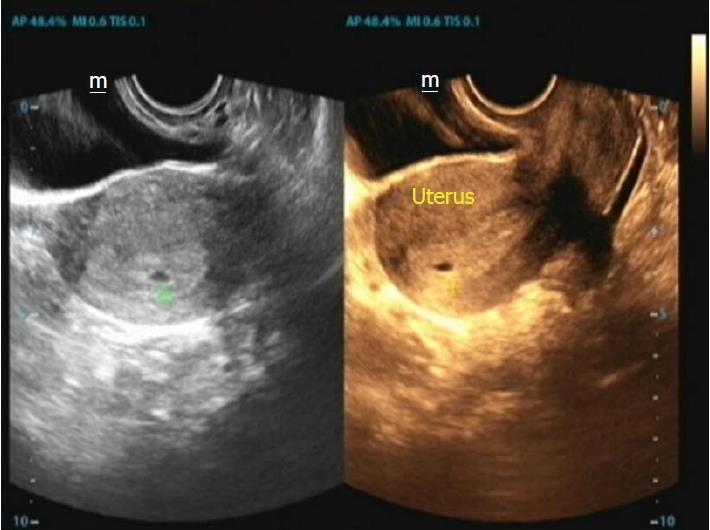
Figure 3 Transvaginal ultrasound after hysteroscopy.
The image shows a 3-mm gestational sac in the uterine cavity.
- Citation: Zhao CY, Ye F. Live birth after hysteroscopy performed inadvertently during early pregnancy: A case report and review of literature. World J Clin Cases 2018; 6(12): 559-563
- URL: https://www.wjgnet.com/2307-8960/full/v6/i12/559.htm
- DOI: https://dx.doi.org/10.12998/wjcc.v6.i12.559


