Copyright
©The Author(s) 2018.
World J Clin Cases. Oct 6, 2018; 6(11): 477-482
Published online Oct 6, 2018. doi: 10.12998/wjcc.v6.i11.477
Published online Oct 6, 2018. doi: 10.12998/wjcc.v6.i11.477
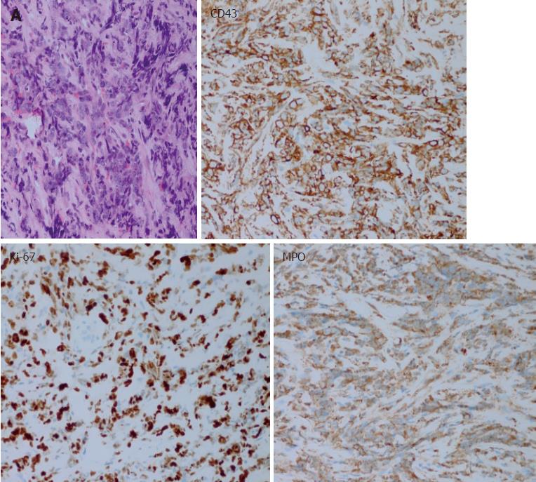
Figure 2 Haematoxylin and eosin stainin (magnification × 400) shows heterotypic cells arranged in a line that appear flaky and demonstrate infiltrative growth (A), the inserts show CD43, Ki-67, and MPO expression.
- Citation: Zhu T, Xi XY, Dong HJ. Isolated myeloid sarcoma in the pancreas and orbit: A case report and review of literature. World J Clin Cases 2018; 6(11): 477-482
- URL: https://www.wjgnet.com/2307-8960/full/v6/i11/477.htm
- DOI: https://dx.doi.org/10.12998/wjcc.v6.i11.477


