Copyright
©The Author(s) 2018.
World J Clin Cases. Oct 6, 2018; 6(11): 472-476
Published online Oct 6, 2018. doi: 10.12998/wjcc.v6.i11.472
Published online Oct 6, 2018. doi: 10.12998/wjcc.v6.i11.472
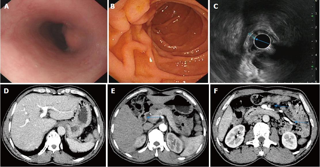
Figure 2 Following administration of praziquantel, the patient with acute Schistosoma japonicum infection received a complete check-up at the follow-up visit.
A, B: Upper gastrointestinal endoscopy showing protrusive lesions of the esophagus (A) and normal mucosa in the descending duodenum (B); C: Endoscopic ultrasonography revealing slight thickening and normal layer of the descending duodenal wall; D-F: Dynamic abdominal computed tomography showing homogeneous hepatic perfusion on the portal phase (D), slight thickening of the descending wall (E, arrow), shrinkage of the swollen mesentery to normal size (F, arrow), and disappearance of ascites.
- Citation: Xiao ZL, Xu KS, Song YH. Unusual cause of lesions in the descending duodenum and liver: A case report and review of literature. World J Clin Cases 2018; 6(11): 472-476
- URL: https://www.wjgnet.com/2307-8960/full/v6/i11/472.htm
- DOI: https://dx.doi.org/10.12998/wjcc.v6.i11.472


