Copyright
©The Author(s) 2018.
World J Clin Cases. Oct 6, 2018; 6(11): 472-476
Published online Oct 6, 2018. doi: 10.12998/wjcc.v6.i11.472
Published online Oct 6, 2018. doi: 10.12998/wjcc.v6.i11.472
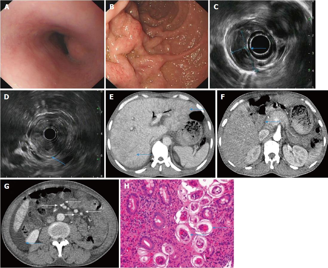
Figure 1 A 47-year-old male patient with acute Schistosoma japonicum infection underwent endoscopic examination, endoscopic ultrasonography and dynamic computed tomography scanning at the baseline visit.
A, B: Upper gastrointestinal endoscopy showing protrusive lesions in the esophagus (A), swollen mucosa and protrusive lesions in the descending duodenum (B); C, D: Endoscopic ultrasonography revealing a hypoechoic mass in the muscularis mucosa of the esophagus (C, arrow), thickening of the descending duodenal wall, and destruction of the descending duodenal wall (D, arrow); E-G: Dynamic computed tomography showing heterogeneous hypointensity in the liver (E, arrow), thickening of the descending duodenal wall (F, arrow), swollen mesentery around the arteries (G, arrow), and ascites; H: Biopsy of the descending duodenum showing deposition of Schistosoma eggs.
- Citation: Xiao ZL, Xu KS, Song YH. Unusual cause of lesions in the descending duodenum and liver: A case report and review of literature. World J Clin Cases 2018; 6(11): 472-476
- URL: https://www.wjgnet.com/2307-8960/full/v6/i11/472.htm
- DOI: https://dx.doi.org/10.12998/wjcc.v6.i11.472


