Copyright
©The Author(s) 2018.
World J Clin Cases. Oct 6, 2018; 6(11): 459-465
Published online Oct 6, 2018. doi: 10.12998/wjcc.v6.i11.459
Published online Oct 6, 2018. doi: 10.12998/wjcc.v6.i11.459
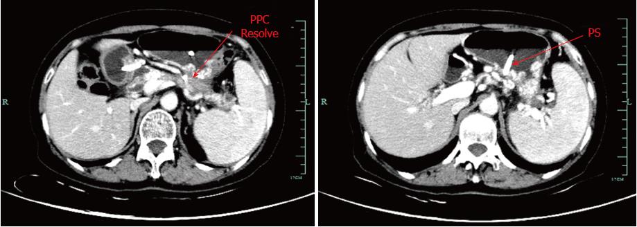
Figure 6 Follow-up contrast-enhanced computed tomography images.
Computed tomography scan of the abdomen revealing a significant decrease in the size of the pseudocyst with a double pig plastic stent in position two months after endoscopy ultrasound-guided placement of a visible plastic stent between the stomach and residual pancreatic pseudocyst. The stent also effectively drained the pseudocyst and relieved the severely affected collateral vessels.
- Citation: Wang BH, Xie LT, Zhao QY, Ying HJ, Jiang TA. Balloon dilator controls massive bleeding during endoscopic ultrasound-guided drainage for pancreatic pseudocyst: A case report and review of literature. World J Clin Cases 2018; 6(11): 459-465
- URL: https://www.wjgnet.com/2307-8960/full/v6/i11/459.htm
- DOI: https://dx.doi.org/10.12998/wjcc.v6.i11.459


