Copyright
©The Author(s) 2018.
World J Clin Cases. Oct 6, 2018; 6(11): 447-454
Published online Oct 6, 2018. doi: 10.12998/wjcc.v6.i11.447
Published online Oct 6, 2018. doi: 10.12998/wjcc.v6.i11.447
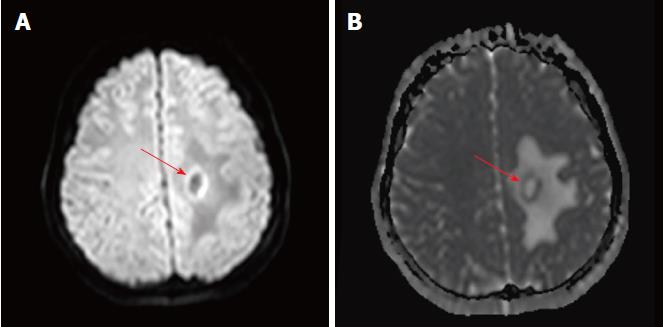
Figure 3 Apparent diffusion co-efficient maps portraying only a thin rim of restricted diffusion at the outer rim of the lesion, with facilitated diffusion centrally and at the outer edema.
A: Diffusion weight images shows a thin rim of increased diffusion at the outer rim of the lesion; B: The outer rim is hypointense on the corresponding apparent diffusion coefficient map images, indicating true restriction.
- Citation: Ertuğrul Ö, Çiçekçi E, Tuncer MC, Aluçlu MU. Balo’s concentric sclerosis in a patient with spontaneous remission based on magnetic resonance imaging: A case report and review of literature. World J Clin Cases 2018; 6(11): 447-454
- URL: https://www.wjgnet.com/2307-8960/full/v6/i11/447.htm
- DOI: https://dx.doi.org/10.12998/wjcc.v6.i11.447


