Copyright
©The Author(s) 2018.
World J Clin Cases. Sep 26, 2018; 6(10): 384-392
Published online Sep 26, 2018. doi: 10.12998/wjcc.v6.i10.384
Published online Sep 26, 2018. doi: 10.12998/wjcc.v6.i10.384
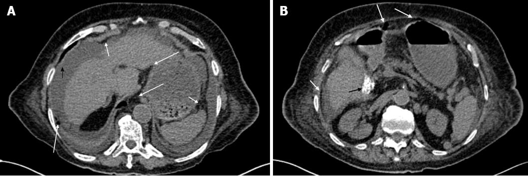
Figure 1 CT scan findings.
A: Evidence of an important abdominopelvic peritoneal effusion (black arrowhead), perigastric free air along the gastrocolic ligament and under the anterior abdominal wall (white arrows), and pericardial and bilateral pleural effusions; B: In addition to perigastric free air and under the abdominal wall (white arrows), a microlithiasis (black arrow) of the gallbladder could also be observed.
- Citation: Fleres F, Ieni A, Saladino E, Speciale G, Aspromonte M, Cannaò A, Macrì A. Rectal perforation by inadvertent ingestion of a blister pack: A case report and review of literature. World J Clin Cases 2018; 6(10): 384-392
- URL: https://www.wjgnet.com/2307-8960/full/v6/i10/384.htm
- DOI: https://dx.doi.org/10.12998/wjcc.v6.i10.384


