Copyright
©The Author(s) 2018.
World J Clin Cases. Sep 26, 2018; 6(10): 344-354
Published online Sep 26, 2018. doi: 10.12998/wjcc.v6.i10.344
Published online Sep 26, 2018. doi: 10.12998/wjcc.v6.i10.344
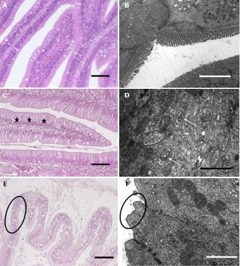
Figure 3 Histopathological and ultrastructural observations in the gastrointestinal tract of Tenca fish exposed to MCs.
Adapted from a ref.[40]. H and E staining (A) and ultrastructural observations (B). A and B: Control groups; C and D: 25 mg of MC-LR/fish group; E and F: 55 mg of MC-LR/fish groups. Vacuolated enterocytes (star). Vacuolization of the endoplasmic reticulum (arrow). Pycnotic nuclei, vacuolated cytoplasm (E, circle). Total microvilli loss (F, circle).
- Citation: Wu JX, Huang H, Yang L, Zhang XF, Zhang SS, Liu HH, Wang YQ, Yuan L, Cheng XM, Zhuang DG, Zhang HZ. Gastrointestinal toxicity induced by microcystins. World J Clin Cases 2018; 6(10): 344-354
- URL: https://www.wjgnet.com/2307-8960/full/v6/i10/344.htm
- DOI: https://dx.doi.org/10.12998/wjcc.v6.i10.344


