Copyright
©The Author(s) 2018.
World J Clin Cases. Sep 26, 2018; 6(10): 322-334
Published online Sep 26, 2018. doi: 10.12998/wjcc.v6.i10.322
Published online Sep 26, 2018. doi: 10.12998/wjcc.v6.i10.322
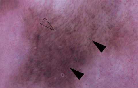
Figure 2 Dermoscopic findings of the Laugier-Hunziker syndrome patient.
Granular (closed arrowhead) and linear (open arrowhead) patterns coexist in the labial pigmented lesion.
- Citation: Duan N, Zhang YH, Wang WM, Wang X. Mystery behind labial and oral melanotic macules: Clinical, dermoscopic and pathological aspects of Laugier-Hunziker syndrome. World J Clin Cases 2018; 6(10): 322-334
- URL: https://www.wjgnet.com/2307-8960/full/v6/i10/322.htm
- DOI: https://dx.doi.org/10.12998/wjcc.v6.i10.322


