Copyright
©The Author(s) 2017.
World J Clin Cases. Aug 16, 2017; 5(8): 344-348
Published online Aug 16, 2017. doi: 10.12998/wjcc.v5.i8.344
Published online Aug 16, 2017. doi: 10.12998/wjcc.v5.i8.344
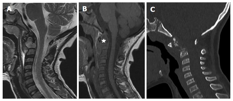
Figure 1 Initial cervical imaging: Sagittal FSET2 (A), SET1 magnetic resonance images (B) and Sagittal thin slice CT image (C).
Infiltrative mass involving the dens of C2 hypointense on T1 and hyperintense on T2 sequence, extending to the surrounding soft tissues (star) leading to an increase in C1-C2 space. No compression of the spinal cervical cord. No signal abnormality nor rupture of the posterior longitudinal ligament spine. Complement CT showed fragmented dens with important C1-C2 dislocation.
- Citation: Tfifha M, Gaha M, Mama N, Yacoubi MT, Abroug S, Jemni H. Atlanto-axial langerhans cell histiocytosis in a child presented as torticollis. World J Clin Cases 2017; 5(8): 344-348
- URL: https://www.wjgnet.com/2307-8960/full/v5/i8/344.htm
- DOI: https://dx.doi.org/10.12998/wjcc.v5.i8.344


