Copyright
©The Author(s) 2017.
World J Clin Cases. Jun 16, 2017; 5(6): 254-257
Published online Jun 16, 2017. doi: 10.12998/wjcc.v5.i6.254
Published online Jun 16, 2017. doi: 10.12998/wjcc.v5.i6.254
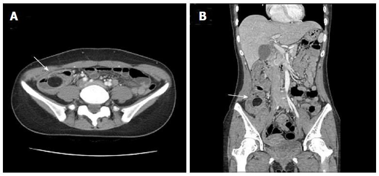
Figure 2 Axial (A) and coronal (B) plain abdominal computed tomography scans demonstrate a well-circumscribed, intraluminal hypodense mass with fat attenuation in the terminal ileum (arrow).
- Citation: Lee DE, Choe JY. Ileocolic intussusception caused by a lipoma in an adult. World J Clin Cases 2017; 5(6): 254-257
- URL: https://www.wjgnet.com/2307-8960/full/v5/i6/254.htm
- DOI: https://dx.doi.org/10.12998/wjcc.v5.i6.254


