Copyright
©The Author(s) 2017.
World J Clin Cases. Jun 16, 2017; 5(6): 222-233
Published online Jun 16, 2017. doi: 10.12998/wjcc.v5.i6.222
Published online Jun 16, 2017. doi: 10.12998/wjcc.v5.i6.222
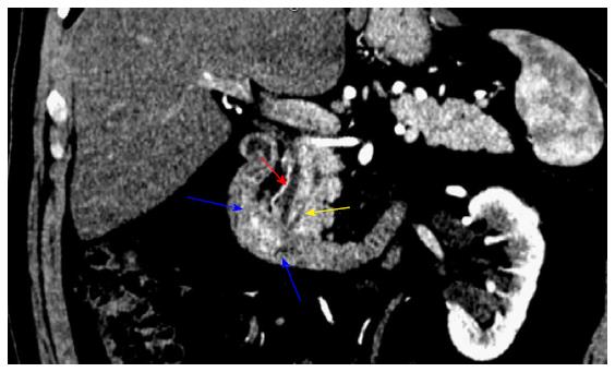
Figure 1 Preoperative computed tomography imaging.
Coronal section demonstrating a 2.1 cm × 1.4 cm periampullary duodenal mass (blue arrows). Red arrow: Common bile duct; yellow arrow: Pancreatic duct.
- Citation: Cathcart SJ, Sasson AR, Kozel JA, Oliveto JM, Ly QP. Duodenal gangliocytic paraganglioma with lymph node metastases: A case report and comparative review of 31 cases. World J Clin Cases 2017; 5(6): 222-233
- URL: https://www.wjgnet.com/2307-8960/full/v5/i6/222.htm
- DOI: https://dx.doi.org/10.12998/wjcc.v5.i6.222


