Copyright
©The Author(s) 2017.
World J Clin Cases. Apr 16, 2017; 5(4): 153-158
Published online Apr 16, 2017. doi: 10.12998/wjcc.v5.i4.153
Published online Apr 16, 2017. doi: 10.12998/wjcc.v5.i4.153
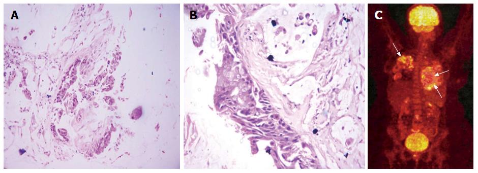
Figure 4 Histopathology and positron emission tomography findings.
A and B: Histological photomicrograph shows fragments of tumor with mucinous epithelium H and E × 40. Higher magnification (C) shows invasive mucinous adenocarcinoma with pools of extracellular mucin H and E × 400; C: Whole-body PET-CT image shows patchy mild uptake of FDG within the lung lesions. No evidence of extrathoracic primary site of malignancy is there. PET-CT: Positron emission tomography-computed tomography; FDG: 2-[fluorine-18] fluoro-2-deoxy-D-glucose.
- Citation: Verma R, Bhalla AS, Goyal A, Jain D, Loganathan N, Guleria R. Ominous lung cavity “Tambourine sign”. World J Clin Cases 2017; 5(4): 153-158
- URL: https://www.wjgnet.com/2307-8960/full/v5/i4/153.htm
- DOI: https://dx.doi.org/10.12998/wjcc.v5.i4.153


