Copyright
©The Author(s) 2017.
World J Clin Cases. Apr 16, 2017; 5(4): 153-158
Published online Apr 16, 2017. doi: 10.12998/wjcc.v5.i4.153
Published online Apr 16, 2017. doi: 10.12998/wjcc.v5.i4.153
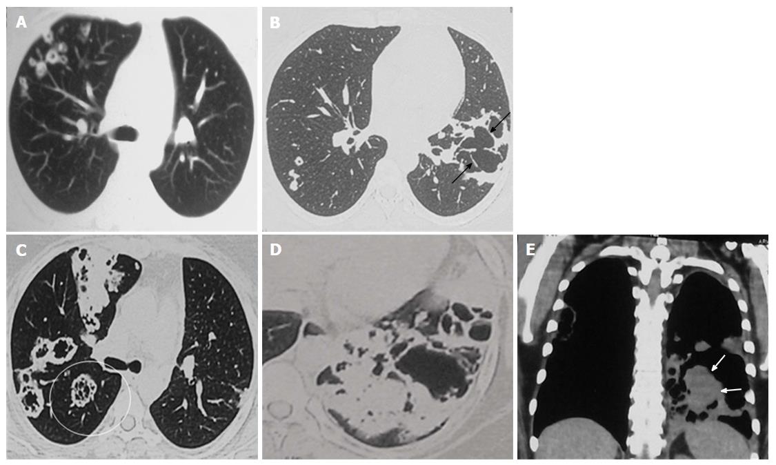
Figure 3 Disease progression with development of soft tissue.
Chest CT study of 2012 lung window (A and B) shows multiple new cavitating nodules in RUL (A) and increase in size of LLL cavity with development of internal septations (arrows) (B). No solid nodules or GGO or consolidation is seen. Current CECT images (2014: C to E) demonstrate further increase in size of the lesions and multiple new lesions having internal septations and development of significant soft tissue component in LLL cavity (solid arrows). Also note the “Tambourine” sign in RLL cavities as well (encircled cavity in C). LLL: Left lower lobe; RUL: Right upper lobe; GGO: Ground glass opacity; CECT: Contrast-enhanced computed tomography.
- Citation: Verma R, Bhalla AS, Goyal A, Jain D, Loganathan N, Guleria R. Ominous lung cavity “Tambourine sign”. World J Clin Cases 2017; 5(4): 153-158
- URL: https://www.wjgnet.com/2307-8960/full/v5/i4/153.htm
- DOI: https://dx.doi.org/10.12998/wjcc.v5.i4.153


