Copyright
©The Author(s) 2017.
World J Clin Cases. Apr 16, 2017; 5(4): 153-158
Published online Apr 16, 2017. doi: 10.12998/wjcc.v5.i4.153
Published online Apr 16, 2017. doi: 10.12998/wjcc.v5.i4.153
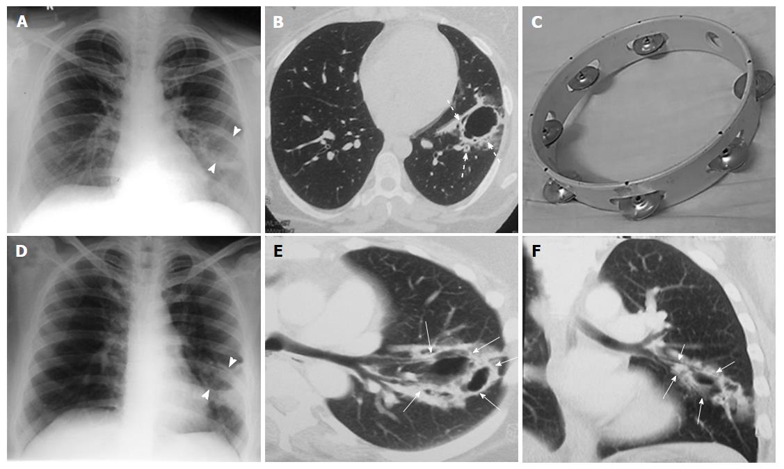
Figure 2 Initial and two year follow-up computed tomography imaging.
Chest radiograph (A) and CT (B) in 2008 (first study) show well-defined thin-walled (4 mm) irregular cavitary lesion (arrow head) in superior segment of left lower lobe. Thick walled bronchioles (dotted arrows) are seen near the edge and within the wall of cavity with adjacent ground glass giving rise to “Tambourine” sign; (C) depicts the musical instrument “tambourine” for comparison; subsequent radiograph (D) and CT (E and F) in 2010 shows increase in size and wall thickness of the cavity. Note the adjacent bronchioles (thin white arrows) entering into the cavity wall. CT: Computed tomography.
- Citation: Verma R, Bhalla AS, Goyal A, Jain D, Loganathan N, Guleria R. Ominous lung cavity “Tambourine sign”. World J Clin Cases 2017; 5(4): 153-158
- URL: https://www.wjgnet.com/2307-8960/full/v5/i4/153.htm
- DOI: https://dx.doi.org/10.12998/wjcc.v5.i4.153


